Review
. 2017 Jun 20:86:485-514.
doi: 10.1146/annurev-biochem-061516-044500.

A New Facet of Vitamin B12: Gene Regulation by Cobalamin-Based Photoreceptors

Affiliations
Review

A New Facet of Vitamin B12: Gene Regulation by Cobalamin-Based Photoreceptors

S Padmanabhan et al. Annu Rev Biochem. .

Abstract

Living organisms sense and respond to light, a crucial environmental factor, using photoreceptors, which rely on bound chromophores such as retinal, flavins, or linear tetrapyrroles for light sensing. The discovery of photoreceptors that sense light using 5'-deoxyadenosylcobalamin, a form of vitamin B12 that is best known as an enzyme cofactor, has expanded the number of known photoreceptor families and unveiled a new biological role of this vitamin. The prototype of these B12-dependent photoreceptors, the transcriptional repressor CarH, is widespread in bacteria and mediates light-dependent gene regulation in a photoprotective cellular response. CarH activity as a transcription factor relies on the modulation of its oligomeric state by 5'-deoxyadenosylcobalamin and light. This review surveys current knowledge about these B12-dependent photoreceptors, their distribution and mode of action, and the structural and photochemical basis of how they orchestrate signal transduction and control gene expression.

Keywords: CarH; chromophore; optogenetics; photochemistry; photoregulation; transcriptional repressor.


Figures

Figure 1
Figure 1
Vitamin B12 and derivatives. (Left) General chemical structure of B12 in the base-on conformation with cobalt formally in the Co(III) state, the lower axial dimethylbenzimidazole ligand in blue, and the upper axial ligand denoted by “R” in red. (Right) Selected upper axial ligands and the corresponding B12 forms are shown.
Figure 2
Figure 2
Experimentally studied CarH and CarA proteins. (a) Genomic context of the carH/carA genes in M. xanthus (Mx), T. thermophilus (Tt) and B. megaterium (Bm). Other genes and their corresponding products are as follows (carotenoid synthesis genes are in blue): crtE (E), farnesyltransferase; crtI (I), phytoene desaturase; crtB (B), phytoene synthase; crtD (D), hydroxyneurosporene dehydrogenase; crtC (C), neurosporene hydroxylase; ?, putative carotenoid biosynthesis protein; crtYc (Yc), crtYd (Yd), components of a heterodimeric lycopene cyclase; white arrow, predicted acyltransferase domain-containing protein; phr, DNA photolyase; ldrP, CRP/FNR family transcriptional regulator. (b) Domain architecture of CarH/CarA proteins. Numbers delimiting the domains are from the crystal structure of CarH from Tt. Characteristic motifs (x, any residue) are shown below for each protein (size in residues is indicated in brackets).
Figure 3
Figure 3
Light-dependent gene regulation mechanisms by B12-binding transcription factors. (a) M. xanthus CarA and CarS. Cooperative binding of CarA dimers to a bipartite operator overlapping the −35 promoter region blocks access to RNA polymerase and represses transcription in the dark. Light induces production of CarS, which sequesters CarA, prevents operator DNA-binding, and activates transcription. (b) M. xanthus/T. thermophilus CarH. CarH monomers in the apo form bind to AdoCbl (filled blue asterisk) to form stable AdoCbl-bound CarH tetramers in the dark, which bind to operator DNA overlapping the −35 promoter region to block access to RNA polymerase and repress transcription. Light disrupts AdoCbl-CarH tetramers to monomers by photolysing bound AdoCbl (unfilled blue asterisks and blue circles correspond to products after photolysis described in the text), loss of operator-binding, and activation of transcription. (c) B. megaterium CarH. ApoCarH tetramers, which do not bind DNA, yield AdoCbl-bound tetramers that bind to operator DNA overlapping the −35 region of PcarH (another operator that overlaps the −10 region of Pcrt is not shown) to repress transcription in the dark. Light disrupts AdoCbl-CarH tetramers to dimers (by photolysing bound AdoCbl), abolishing operator binding and relieving repression. (d) R. capsulatus CrtJ and AerR. CrtJ dimers repress transcription in the dark by binding to two sites overlapping the −10 and −35 promoter regions. On exposure to light, binding of AqCbl (produced by AdoCbl or MeCbl photolysis) to AerR enables its association with CrtJ to disrupt CrtJ dimers and DNA-binding, activating transcription.
Figure 4
Figure 4
Dark state AdoCbl-bound CarH compared to structurally similar DNA- and B12-binding domains. (a) CarH protomer structure [Protein Data Bank (PDB) accession code 5C8D] showing the modules for DNA-binding (compared with that in CarA, top left; PDB accession code 2JML) and B12-binding (compared to that in MetH, bottom left; PDB accession code 1BMT). (b) Close-up of the B12-binding site showing residues contacting the upper axial ligand (in cyan) of MeCbl in MetH and cobalt-coordinating His (H759). (c) Close-up of the B12-binding site showing residues contacting the upper axial ligand (in cyan) of AdoCbl in CarH and cobalt-coordinating His (H177). For a better view, the orientation in b and c is slightly different from that in a. (d) Comparison (in stereo) of the orientations of the AdoCbl Ado group in CarH, in free AdoCbl, and in selected enzymes whose structures have been determined with intact AdoCbl: IcmF (PDB accession code 4XC6) and related mutases; ornithine-aminomutase (OAM) in its resting state (PDB accession code 3KP1).
Figure 5
Figure 5
The CarH tetramer and its unexpected DNA binding mode. (a) The head-to-tail packing of the two protomers in a CarH dimer, with the helix bundle colored yellow, the AdoCbl-binding domain in green, and the two DBDs in cyan. On the right is a close-up of the extensive interface of the head-to-tail dimer with several H-bonds and ionic interactions indicated. D201 and R176 indicate residues whose interaction was shown to be crucial by mutational analysis. (b) The CarH tetramer of two head-to-tail dimers with the four DBDs (in purple, cyan, light cyan, and dark blue) [Protein Data Bank (PDB) accession code 5C8D]. Two alternative views of the tetramer are shown to better appreciate the complex quaternary structure of CarH. The view on the left shows the distribution of the DBDs on the protein surface (DBDs shown in pink and dark blue correspond to the head-to-tail dimer at the back, whose B12-binding domains are shown in gray). (c) CarH tetramer (colored as in panel b) in complex with DNA (PDB accession code 5C8E) is shown. Three DBDs are reoriented and contact three direct repeats (DR) in the DNA sequence, depicted schematically below (structures in b and d are redrawn versions of those previously reported in (38)). (d) Direct repeats recognized by CarH at PcarH in T. thermophilus (Tt) determined from the structure of the complex, and those inferred in M. xanthus (Mx) and B. megaterium (Bm) from footprinting data and sequence inspection. Base pairs covered by the recognition helix, as deduced from the CarH-DNA structure, are shaded. The −35 promoter region is shown in red. In Mx PB, the two CarA binding site palindromes are underlined. The repeats in Bm Pcrt and its −10 region (in purple) correspond to the noncoding strand. The DNA sequence logo for a probable consensus CarH direct repeat recognition site from the sequences above is shown (bottom).
Figure 6
Figure 6
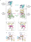
Molecular basis of photoregulation by CarH. (a) Structure of light-exposed CarH [solid, Protein Data Bank (PDB) accession code 5C8F] with the arrow indicating the major shift in the helix bundle relative to the dark structure (transparent, PDB accession code 5C8D). (b) Close-up of the AdoCbl-binding site highlighting the role of the upper axial Ado group (in cyan) of AdoCbl as a molecular doorstop in the dark state. The arrow indicates the helix bundle shift that occurs on exposure to light leading to relocation of W131 and the other indicated residues. The nonconserved E129 was suggested to be involved in forming the bis-His adduct by deprotonating His132 based on homology modeling and molecular dynamics studies (40), but this may be unlikely given its positioning in the crystal structure. (c) Close-up of the bis-His Co coordination in light-exposed CarH. (d) Scheme of the proposed mechanism for CarH photolysis adapted from (39), with additional details from the high-resolution structures (38) and apparent rates, kapp, from transient kinetics data (40). The chemical structure of 4′,5′-anhydroadenosine, the product of AdoCbl photolysis in CarH (39), is shown in the middle (and to the right) of the scheme.
Figure 7
Figure 7
Phylogenetic distribution of CarH and of its B12-binding domain in other proteins. (a) Distribution of the indicated domain architectures across different bacterial phyla, with the number of genomes for each phylum indicated (from ~8400 bacterial genomes available at http://www.ncbi.nlm.nih.gov/genomes; for simplicity, only Candidatus phyla with relevant B12-binding domains are shown). The number of proteins in each phylum (H, CarH; A, CarA) correspond to the color heat map on the bottom right. InterPro (protein sequence analysis & classification, https://www.ebi.ac.uk/interpro/) was used to search for proteins with a B12-binding domain (IPR006158) alone, or in combination with a MerR-type DNA-binding (IPR000551) domain or other domains [IPR007024, globin-like; IPR005467, histidine kinase; IPR029016, GAF (cGMP-specific phosphodiesterases, adenylyl cyclase, FhlA)]. Protein sequences retrieved from UniProt (Universal Protein Resource, http://www.uniprot.org/) were aligned using MUSCLE in MEGA7 (Molecular Evolutionary Genetics Analysis, http://www.megasoftware.net/). Those with the E/DxH B12-binding motif preceded by a W(9/10)xEH motif were selected by manual curation. The ones fused to a MerR-type DBD with the RxWxxR motif were classified as CarH or, if they lacked the Trp but not the EH of the W(9/10)xEH motif, as CarA. The 200-260 residue size range was used to select for standalone proteins. (b) Sequence logo created using WebLogo (http://weblogo.berkeley.edu/) for 498 CarH proteins with the W-(9)x-EH motif (those with a W-(10)x-EH motif, mostly in actinobacteria, were omitted). Shown are segments around signature residues (asterisks above) used for curation in the DBD (top left, cyan border), the four-helix bundle (top right, gold border) and the Rossmann fold (bottom, green border). In the sequences (x, any residue) below each logo, conserved residues (≥95%) are highlighted in bold and larger font.

Similar articles

Cited by

References

    1. Croce R, van Amerongen H. 2014. Natural strategies for photosynthetic light harvesting. Nat. Chem. Biol 10: 492–501 - PubMed
    1. Palczewski K. 2012. Chemistry and biology of vision. J. Biol. Chem 287: 1612–19 - PMC - PubMed
    1. Wald G. 1968. The molecular basis of visual excitation. Nature 219: 800–07 - PubMed
    1. Cohen SE, Golden SS. 2015. Circadian rhythms in cyanobacteria. Microbiol. Mol. Biol. Rev 79: 373–85 - PMC - PubMed
    1. Nagel DH, Kay SA. 2012. Complexity in the wiring and regulation of plant circadian networks. Curr. Biol 22: R648–57 - PMC - PubMed

Publication types

MeSH terms

LinkOut - more resources