

Image-Based Analysis of Tumor Localization After Intra-Arterial Delivery of Technetium-99m-Labeled SPIO Using SPECT/CT and MRI
- PMID: 28654377
- PMCID: PMC5470132
- DOI: 10.1177/1536012116689001
Image-Based Analysis of Tumor Localization After Intra-Arterial Delivery of Technetium-99m-Labeled SPIO Using SPECT/CT and MRI
Abstract
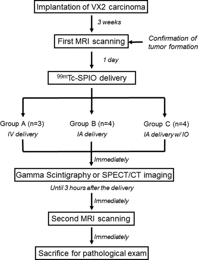
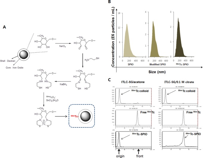
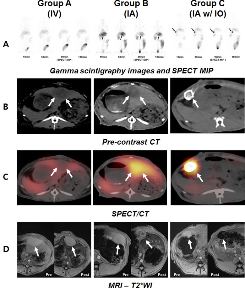
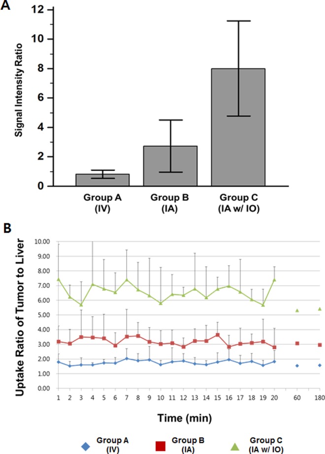
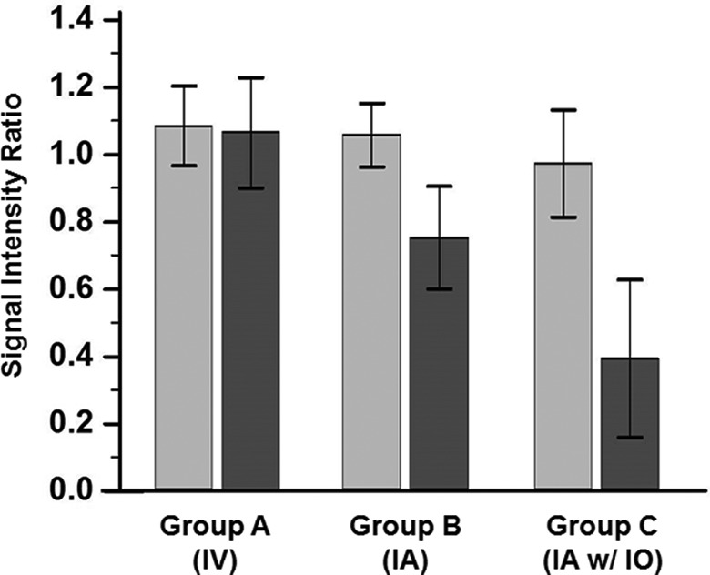
The aim of this study is to evaluate the localization of 99mTc-labeled dextran-coated superparamagnetic iron oxide (SPIO) nanoparticles to the liver tumor using image-based analysis. We delivered 99mTc-SPIO intravenously or intra-arterially (IA) with/without Lipiodol to compare the tumor localization by gamma scintigraphy, single-photon emission computed tomography (SPECT), and magnetic resonance imaging (MRI) in a rabbit liver tumor. The gamma and SPECT image-based analysis shows that the uptake ratio of the tumor to the normal liver parenchyma is highest after delivery of 99mTc-SPIO with Lipiodol IA and that well correlates with the trend of the signal decrease in the liver MRIs. Intra-arterial delivery of SPIO with Lipiodol might be a good drug delivery system targeting the hepatic tumors, as confirmed by image-based analysis.
Keywords: Intra-arterial (IA) delivery; magnetic resonance imaging (MRI); single-photon emission computed tomography (SPECT); superparamagnetic iron oxide (SPIO); technetium-99m.
Conflict of interest statement
Figures
Similar articles
-
99mTc-Diethylenetriamine pentaacetic acid superparamagnetic iron oxide nanoparticles conjugated with lactobionic acid.2010 Apr 28 [updated 2010 May 20]. In: Molecular Imaging and Contrast Agent Database (MICAD) [Internet]. Bethesda (MD): National Center for Biotechnology Information (US); 2004–2013. 2010 Apr 28 [updated 2010 May 20]. In: Molecular Imaging and Contrast Agent Database (MICAD) [Internet]. Bethesda (MD): National Center for Biotechnology Information (US); 2004–2013. PMID: 20641996 Free Books & Documents. Review.
-
Comparison between contrast-enhanced magnetic resonance imaging and technetium 99m glucohepatonic acid single photon emission computed tomography with histopathologic correlation in gliomas.J Comput Assist Tomogr. 2006 Sep-Oct;30(5):723-33. doi: 10.1097/01.rct.0000228154.58281.88. J Comput Assist Tomogr. 2006. PMID: 16954918 Clinical Trial.
-
99mTc-labeled superparamagnetic iron oxide nanoparticles for multimodality SPECT/MRI of sentinel lymph nodes.J Nucl Med. 2012 Mar;53(3):459-63. doi: 10.2967/jnumed.111.092437. Epub 2012 Feb 9. J Nucl Med. 2012. PMID: 22323777
-
Quantitative analysis of liver function using superparamagnetic iron oxide- and Gd-EOB-DTPA-enhanced MRI: comparison with Technetium-99m galactosyl serum albumin scintigraphy.Eur J Radiol. 2012 Jun;81(6):1100-4. doi: 10.1016/j.ejrad.2011.02.053. Epub 2011 Mar 23. Eur J Radiol. 2012. PMID: 21435811
-
Intra-Arterial Delivery of Cell Therapies for Stroke.Stroke. 2018 May;49(5):1075-1082. doi: 10.1161/STROKEAHA.117.018288. Epub 2018 Apr 18. Stroke. 2018. PMID: 29669876 Free PMC article. Review. No abstract available.
Cited by
-
Biodistribution of Mesoporous Carbon Nanoparticles via Technetium-99m Radiolabelling after Oral Administration to Mice.Nanomaterials (Basel). 2021 Nov 30;11(12):3260. doi: 10.3390/nano11123260. Nanomaterials (Basel). 2021. PMID: 34947611 Free PMC article.
-
Advances in multimodal imaging techniques in nanomedicine: enhancing drug delivery precision.RSC Adv. 2025 Jul 30;15(33):27187-27209. doi: 10.1039/d5ra03255e. eCollection 2025 Jul 25. RSC Adv. 2025. PMID: 40740223 Free PMC article. Review.
-
99mTc-Labeled Iron Oxide Nanoparticles as Dual-Modality Contrast Agent: A Preliminary Study from Synthesis to Magnetic Resonance and Gamma-Camera Imaging in Mice Models.Nanomaterials (Basel). 2022 Aug 8;12(15):2728. doi: 10.3390/nano12152728. Nanomaterials (Basel). 2022. PMID: 35957159 Free PMC article.
-
Biologically Targeted Magnetic Hyperthermia: Potential and Limitations.Front Pharmacol. 2018 Aug 2;9:831. doi: 10.3389/fphar.2018.00831. eCollection 2018. Front Pharmacol. 2018. PMID: 30116191 Free PMC article. Review.
References
-
- Jemal A, Bray F, Center MM, Ferlay J, Ward E, Forman D. Global cancer statistics. CA Cancer J Clin. 2011;61(2):69–90. - PubMed
-
- Kim YI, Park JW, Kwak HW, et al. Long-term outcomes of second treatment after initial transarterial chemoembolization in patients with hepatocellular carcinoma. Liver Int. 2014;34(8):1278–1286. - PubMed
-
- Bruix J, Sherman M. Management of hepatocellular carcinoma. Hepatology. 2005;42(5):1208–1236. - PubMed
-
- Forner A, Llovet JM, Bruix J. Hepatocellular carcinoma. Lancet. 2012;379(9822):1245–1255. - PubMed
Publication types
MeSH terms
Substances
LinkOut - more resources
Full Text Sources
Other Literature Sources
Medical
