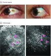
Handheld In Vivo Reflectance Confocal Microscopy for the Diagnosis of Eyelid Margin and Conjunctival Tumors
- PMID: 28654937
- PMCID: PMC5710297
- DOI: 10.1001/jamaophthalmol.2017.2019
Handheld In Vivo Reflectance Confocal Microscopy for the Diagnosis of Eyelid Margin and Conjunctival Tumors
Abstract
Importance: The clinical diagnosis of conjunctival and eyelid margin tumors is challenging, and new noninvasive imaging techniques could be valuable in this field.
Objective: To assess the diagnostic accuracy of handheld in vivo reflectance confocal microscopy (IVCM) for the diagnosis of eyelid margin and conjunctival tumors.
Design: A prospective observational study was conducted at University Hospital of Saint-Etienne from January 2, 2011, to December 31, 2016 (inclusion of patients until December 31, 2015, and follow-up until December 31, 2016). A total of 278 consecutive patients with eyelid margin or conjunctival lesions were included. Conjunctival lesions were diagnosed with a conventional clinical examination using a slitlamp and by handheld IVCM. Final diagnoses were established by histopathologic examination for 155 neoformations suspicious for being malignant through clinical and/or IVCM examination that were excised and on follow-up of 12 months or longer for the remaining 140 lesions.
Main outcomes and measures: Sensitivity, specificity, and positive and negative predictive values for malignant tumors of the conjunctiva and eyelid margin were calculated using clinical examination with slitlamp and handheld IVCM.
Results: In the 278 patients (136 [48.9%] females; mean [SD] age, 59 [21] years), a total of 166 eyelid margin and 129 conjunctival lesions were included in the analysis. Of the 155 excised neoformations with a histopathologic diagnosis, IVCM showed higher sensitivity compared with clinical examination conducted with the slitlamp for malignant tumors of the eyelid margin (98% vs 92%) and conjunctiva (100% vs 88%). The specificity for malignant eyelid margin tumors was higher for IVCM than for slitlamp examination (74% vs 46%), but slightly less for malignant conjunctival tumors (78% vs 88%). Analysis of all neoformations (155 excised and 140 in follow-up) confirmed these differences in the diagnostic accuracy of the clinical examination and IVCM. The presence of hyperreflective Langerhans cells mimicking malignant melanocytes was the main cause for misdiagnosis of malignant conjunctival tumors with IVCM.
Conclusions and relevance: Handheld IVCM could be a useful tool for the identification of malignant conjunctival tumors. Further studies are required to confirm the usefulness of this device and identify possible features that can differentiate Langerhans cells from malignant melanocytes to prevent the misdiagnosis of melanoma using IVCM.
Conflict of interest statement
Figures


Comment in
-
Evolving Technologies for Lid and Ocular Surface Neoplasias: Is Optical Biopsy a Reality?JAMA Ophthalmol. 2017 Aug 1;135(8):852-853. doi: 10.1001/jamaophthalmol.2017.2009. JAMA Ophthalmol. 2017. PMID: 28655031 No abstract available.
References
-
- Cinotti E, Perrot JL, Labeille B, et al. . In vivo confocal microscopy for eyelids and ocular surface: a new horizon for dermatologists. G Ital Dermatol Venereol. 2015;150(1):127-129. - PubMed
-
- Cinotti E, Labeille B, Cambazard F, Thuret G, Gain P, Perrot JL. Reflectance confocal microscopy for mucosal diseases. G Ital Dermatol Venereol. 2015;150(5):585-593. - PubMed
-
- Cinotti E, Perrot J-L, Labeille B, et al. . Handheld reflectance confocal microscopy for the diagnosis of conjunctival tumors. Am J Ophthalmol. 2015;159(2):324-33.e1. - PubMed
-
- Kaspi M, Habougit C, Grivet D, et al. . [The role of reflectance confocal microscopy and optical coherence tomography in the diagnosis of epithelial-cystic conjunctival nevus]. Ann Dermatol Venereol. 2016;143(10):653-656. - PubMed
Publication types
MeSH terms
LinkOut - more resources
Full Text Sources
Other Literature Sources

