. 2017 Jun 27;14(1):131.
doi: 10.1186/s12974-017-0901-y.

Protease-activated receptor-1 activation by granzyme B causes neurotoxicity that is augmented by interleukin-1β

Affiliations

Protease-activated receptor-1 activation by granzyme B causes neurotoxicity that is augmented by interleukin-1β

Paul R Lee et al. J Neuroinflammation. .

Abstract

Background: The cause of neurodegeneration in progressive forms of multiple sclerosis is unknown. We investigated the impact of specific neuroinflammatory markers on human neurons to identify potential therapeutic targets for neuroprotection against chronic inflammation.

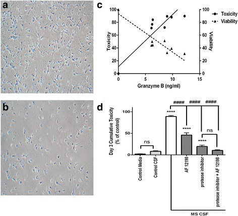
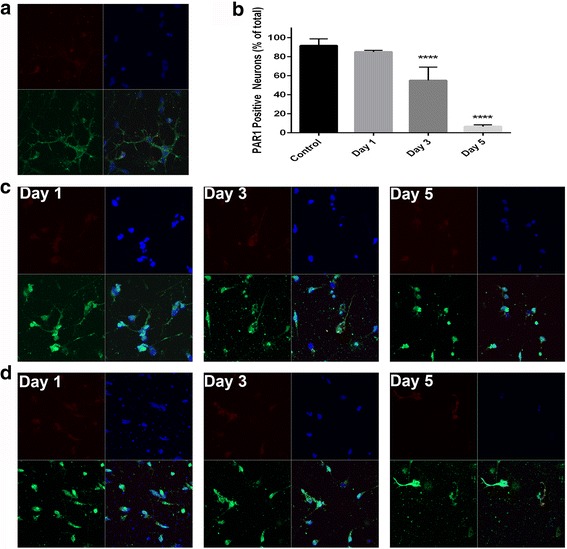
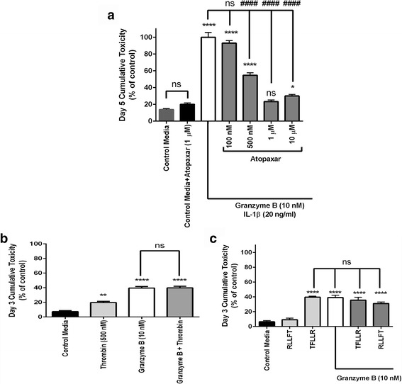
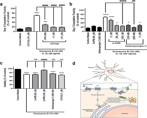
Methods: Surface immunocytochemistry directly visualized protease-activated receptor-1 (PAR1) and interleukin-1 (IL-1) receptors on neurons in human postmortem cortex in patients with and without neuroinflammatory lesions. Viability of cultured neurons was determined after exposure to cerebrospinal fluid from patients with progressive multiple sclerosis or purified granzyme B and IL-1β. Inhibitors of PAR1 activation and of PAR1-associated second messenger signaling were used to elucidate a mechanism of neurotoxicity.

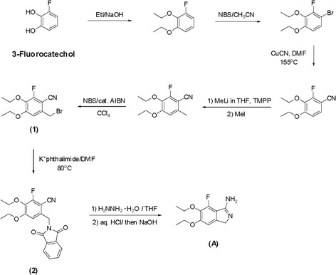
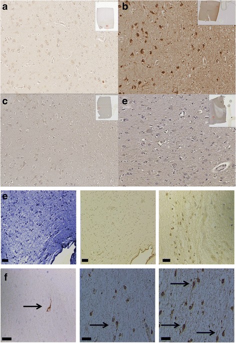
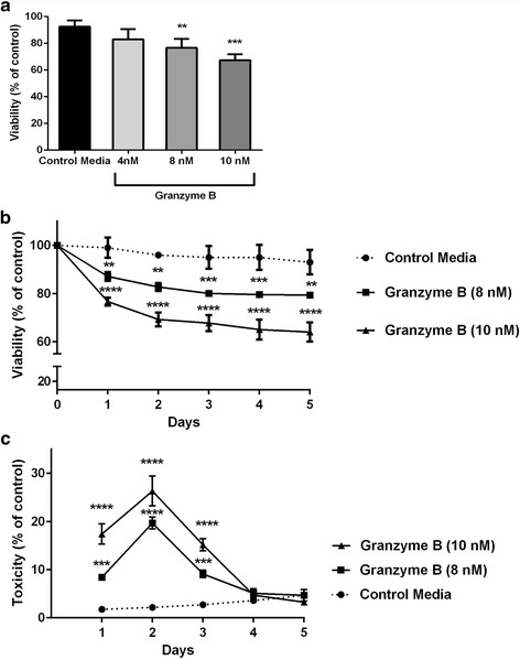
Results: Immunohistochemistry of human post-mortem brain tissue demonstrated cells expressing higher amounts of PAR1 near and within subcortical lesions in patients with multiple sclerosis compared to control tissue. Human cerebrospinal fluid samples containing granzyme B and IL-1β were toxic to human neuronal cultures. Granzyme B was neurotoxic through activation of PAR1 and subsequently the phospholipase Cβ-IP3 second messenger system. Inhibition of PAR1 or IP3 prevented granzyme B toxicity. IL-1β enhanced granzyme B-mediated neurotoxicity by increasing PAR1 expression.

Conclusions: Neurons within the inflamed central nervous system are imperiled because they express more PAR1 and are exposed to a neurotoxic combination of both granzyme B and IL-1β. The effects of these inflammatory mediators may be a contributing factor in the progressive brain atrophy associated with neuroinflammatory diseases. Knowledge of how exposure to IL-1β and granzyme B act synergistically to cause neuronal death yields potential novel neuroprotective treatments for neuroinflammatory diseases.

Keywords: Granzyme B; Interleukin-1; Multiple sclerosis; Neuroinflammation; Neurotoxicity; Protease-activated receptor.


Figures

Scheme 1
Scheme 1
Synthesis of florinated cyclic benzamidine
Scheme 2
Scheme 2
Synthesis of morpholine-substituted phenacyl bromide
Fig. 1
Fig. 1
PAR1 and IL-1 receptor surface expression in human brain tissue. a Immunohistochemistry for PAR1 demonstrates a low level of baseline staining in healthy human brain tissue. b There is more prominent PAR1 staining within MS brain tissue. c IL-1 receptor staining in healthy tissue and d around a demyelinated plaque were similar. e Luxol fast blue stain shows a demyelinated region and subsequent images show PAR1 staining is pronounced within a lesion’s borders and follows a gradient diminishing at approach to the lesion’s outermost border. f Within demyelinated lesions there are darkly stained PAR1-positive neurons with visible unmyelinated axons (arrows). Bars = 50 μm
Fig. 2
Fig. 2
Effects of cerebrospinal fluid (CSF) samples from patients with MS on human neuronal cultures. Cortical neurons derived from induced pluripotent stem cells were treated with undiluted human CSF from patients with MS or from patients without neuroinflammatory diseases for 3 days. a Neuron cultures maintained in CSF from control patients verified to have low levels of granzyme B and interleukin-1β had no evidence of injury or toxicity. b Neuron cultures exposed to CSF from patients with MS were fewer in number and had morphological evidence of damage (i.e., shorter axons, simplified arborization). c The toxicity induced by CSF from patients with MS was directly related to the CSF’s granzyme B content. Conversely, neuronal viability diminished with increasing concentration of granzyme B within a given sample. Data are shown as the mean from duplicate experiments. d The neurotoxicity of CSF from patients with MS was significantly greater at 3 days than CSF from control patients and could be abolished by the cell-permeable granzyme B inhibitor Z-Ala-Ala-Asp-chloromethylketone (25 μM) and partially prevented by inhibition of the interleukin-1 receptor using the antagonist AF 12198 (1 μM). A mixture of granzyme B inhibitor and AF 12198 was no more effective than the granzyme B inhibitor alone at preventing cumulative neurotoxicity. ****p < 0.0001 relative to CSF from control patients, ####p < 0.001 relative to CSF from patients with MS, one-way ANOVA with post hoc Tukey’s test, n = 7 per condition. Data are shown as the mean ± standard error of the mean from duplicate experiments
Fig. 3
Fig. 3
Granzyme B-mediated neurotoxicity. a Pure cortical neuron cultures were treated with granzyme B and the viability and toxicity were measured simultaneously. A single, 24-h exposure to granzyme B at escalating concentrations (4, 8, and 10 nM) replicated a dose-dependent decrease in neuron viability. The 8- and 10-nM doses caused significantly decreased viability relative to control media. **p < 0.01 and ***p < 0.001, respectively, one-way ANOVA with post hoc Dunnett test, n = 8 per condition. Data are shown as the mean ± standard error of the mean from four separate experiments. b When granzyme B was added for five consecutive days, the viability of the all cultures stabilized at day 3. **p < 0.01, ***p < 0.001 and ****p < 0.0001 relative to control media, repeated measures ANOVA with post hoc Dunnett test, n = 10 per condition. Data are shown as the mean ± standard error of the mean from three separate experiments. c Granzyme B-mediated toxicity peaked on day 2 of exposure followed by a decline to control toxicity levels on day 4. ***p < 0.001 and ****p < 0.0001 relative to control media, repeated measures ANOVA post hoc Dunnett test, n = 10 per condition. Data are shown as the mean ± standard error of the mean from three separate experiments
Fig. 4
Fig. 4
PAR1 and IL-1 receptor surface expression on cultured neurons. a At baseline on day 0, most neurons showed surface expression of PAR1 by immunocytochemistry visualized here using a green fluorescent secondary antibody (bottom left panels). A red fluorescent antibody was used to visualize IL-1 receptor (top left panels), and IL-1 receptor surface expression was noted on all neurons and was not significantly changed during granzyme B exposure. Nuclei were stained with DAPI (blue) and superimposed onto the combined image (lower right panels). b The percentage of neurons with PAR1 surface expression visible by fluorescence significantly decreased over the course of 5 days of chronic exposure to granzyme B and IL-1β relative to the total number of DAPI-stained nuclei at any given time ****p < 0.0001 relative to control media, repeated measures ANOVA with post hoc Dunnett test, n = 8 per condition. Data are shown as mean ± standard error of the mean from four separate experiments. c On days 1, 3, and 5 in media containing granzyme B (10 nM), there was a reduction in the number of neurons in culture evident at all time points and a morphological changes consistent with chronic injury including shortened axons. d After 1, 3, and 5 days in media containing granzyme B (10 nM) and IL-1β (20 ng/ml), the total number of neurons in culture, and the number of neurons staining positive for PAR1 were both significantly reduced
Fig. 5
Fig. 5
Enhancement of granzyme B toxicity with IL-1β. a IL-1β (20 ng/ml) augments granzyme B neurotoxicity. Cumulative neurotoxicity of combined granzyme B and IL-1β reaches 100%. ****p < 0.0001 cumulative toxicity relative to control media, ####p < 0.0001 cumulative toxicity relative to granzyme B + IL-1β (two-way ANOVA with post hoc Tukey’s test, n = 5/condition. Data are mean ± standard error of the mean from three separate experiments). b IL-1β is not toxic by itself. The IL-1 receptor antagonist AF 12198 (1 μM) abolished IL-1β effect after 3 days of co-culture with granzyme B and IL-1β (20 ng/ml). ****p < 0.0001 relative to control media, ####p < 0.0001 between granzyme B + IL-1β and granzyme B+ IL-1β + IL-1 receptor antagonist, repeated measures ANOVA with post hoc Tukey’s test, n = 6/condition. Data are mean ± standard error of the mean from three separate experiments. c After 3 days of exposure, IL-1β (20 ng/ml) significantly increased PAR1 mRNA as shown by qPCR and PAR1 protein relative to control levels using Western blot analysis. The IL-1 receptor antagonist AF 12198 (1 μM) blocked the IL-1β effect on PAR1 mRNA and protein. A pooled PAR1 siRNA preparation reduced PAR1 mRNA and protein below control conditions. d Western blot analysis (below bar graph) confirmed the relative amounts of PAR1 protein in respective conditions. *p < 0.05, ****p < 0.0001 relative to control media, one-way ANOVA with post hoc Tukey’s test, n = 4/condition. Data are mean ± standard error of the mean from duplicate experiments. e Granzyme B had no toxicity when PAR1 siRNA reduced PAR1 protein levels. A pooled PAR1 siRNA preparation inhibited all granzyme B neurotoxicity; IL-1β had no effect. Two control siRNA, anti-GAPDH, and a scrambled PAR1 sequence had no effect. ****p < 0.0001 relative to control media, ####p < 0.0001 between PAR1 siRNA + granzyme B+ IL-1β and other siRNA as noted, one-way ANOVA with post hoc Tukey’s test, n = 8/condition. Data are mean ± standard error of the mean from four separate experiments
Fig. 6
Fig. 6
Effects of PAR1 antagonism and agonism. a Atopaxar, a selective antagonist of PAR1 significantly reduced granzyme B total cumulative toxicity at 500 nM, 1 μM, and 10 μM pre-treatment doses after 5 days of co-incubation with granzyme B and IL-1β. The cumulative toxicity at the 1 μM dose of Atopaxar was not significantly different from toxicity of control media, which contained the same concentration of DMSO necessary to solubilize Atopaxar, nor was Atopaxar itself significantly more toxic than control media. *p < 0.05, ****p < 0.0001 relative to control media, ####p < 0.0001 relative to granzyme B+ IL-1β, repeated measures ANOVA with post hoc Tukey’s test, n = 8 per condition. Data are shown as the mean ± standard error of the mean from five separate experiments. b Thrombin (500 nM), an agonist of PAR1, causes mild neurotoxicity when added to neuronal cultures for 3 days. When granzyme B is added with thrombin, there is no additive toxic effect. *p < 0.05, ****p < 0.0001 relative to control media, one-way ANOVA with post hoc Tukey’s test, n = 10 per condition. Data are shown as the mean ± standard error of the mean from duplicate experiments. c The peptide activating sequence TFLLR (5 μM), an agonist of PAR1, reduced neuronal viability after 3 days to the same extent as 10 nM granzyme B. When granzyme B and TFLLR were combined, there was no additional increase in toxicity. RLLFT, the inverse peptide of the agonist sequence, was non-toxic by itself and did not increase toxicity when paired with granzyme B. ****p < 0.0001 relative to control media, one-way ANOVA with post hoc Tukey’s test, n = 10 per condition. Data are shown as the mean ± standard error of the mean from three separate experiments
Fig. 7
Fig. 7
Inhibition of protein kinase C and IP3 reduced neurotoxicity. a Pre-treatment with the phospholipase C inhibitor U73122 for 30 min significantly reduced neurotoxicity caused by 3 days of exposure to granzyme B (10 nM) and IL-1β (20 ng/ml). All doses of U73122 were effective at reducing toxicity relative to maximum IL-1β augmented granzyme B-mediated toxicity. The 1 and 10 μM doses reduced toxicity to levels identical to that of control media. ***p < 0.001, ****p < 0.0001 relative to control media with 10% ethanol (vehicle), ####p < 0.0001 relative to granzyme B + IL-1β condition, one-way ANOVA with post hoc Tukey’s test, n = 5 per condition. Data are shown as the mean ± standard error of the mean from five separate experiments. b Several doses of the selective IP3 inhibitors 2-APB and xestospongin significantly reduced the cumulative toxicity typically seen after 3 days. *p < 0.05, ****p < 0.0001 relative to control media and ##p < 0.01, ####p < 0.0001 relative to granzyme B + IL-1β, one-way ANOVA with post hoc Tukey’s test, n = 5 per condition. Data are shown as the mean ± standard error of the mean from four separate experiments. c Neuronal viability was preserved in an acute, single day exposure to 10 nM granzyme B following pre-treatment with only 100 μM 2-APB, ****p < 0.0001 relative to control media, ####p < 0.0001 relative to granzyme B + IL-1β, one-way ANOVA with post hoc Tukey’s test, n = 5 per condition. Data are shown as the mean ± standard error of the mean from duplicate experiments. d Proposed mechanism of action for granzyme B-mediated neurotoxicity with IL-1β assistance. IL-1β promotes increased numbers of PAR1 receptors on neuronal membrane surface. Granzyme B cleaves the PAR1 receptors’ extracellular domains. Activated PAR1 directly couples with phospholipase Cβ which in turn yields increased IP3 production and leads to the well-described cellular processes that compromise mitochondrial function and eventually culminate in neuronal demise
Fig. 8
Fig. 8
Effect of other second messengers on neurotoxicity. a Pre-treatment with four different inhibitors (Ro 32–0432 hydrochloride, LY 294002, FR 180204, Src Inhibitor-1) directed selectively against second messengers (protein kinase C, PI3, ERK, and src kinase, respectively) all of which activated by PAR1 failed to achieve significant reduction in cumulative toxicity after 3 days of repeated exposure to granzyme B and IL-1β. *p < 0.05, ****p < 0.0001 relative to control media, ##p < 0.01, ####p < 0.0001 relative to granzyme B + IL-1β condition, repeated measures ANOVA with post hoc Tukey’s test, n = 6 per condition. Data are shown as the mean ± standard error of the mean from four separate experiments. b 100 μM Minocycline produced a modest reduction (<20%) in IL-1β augmented granzyme B neurotoxicity. #p < 0.05 relative to granzyme B + IL-1β condition, repeated measures ANOVA with post hoc Tukey’s test, n = 5 per condition. Data are shown as the mean ± standard error of the mean from four separate experiments

References

    1. Anderson DW, Ellenberg JH, Leventhal CM, et al. Revised estimate of the prevalence of multiple sclerosis in the United States. Ann Neurol. 1992;31:333–336. doi: 10.1002/ana.410310317. - DOI - PubMed
    1. Rissanen E, Tuisku J, Rokka J, et al. In Vivo Detection of Diffuse Inflammation in Secondary Progressive Multiple Sclerosis Using PET Imaging and the Radioligand 11C-PK11195. J Nucl Med. 2014;55:939–944. doi: 10.2967/jnumed.113.131698. - DOI - PubMed
    1. Popescu V, Agosta F, Hulst HE, et al. Brain atrophy and lesion load predict long term disability in multiple sclerosis. J Neurol Neurosurg Psychiatry. 2013;84:1082–1091. doi: 10.1136/jnnp-2012-304094. - DOI - PubMed
    1. Fernández-Jaén A, Fernández-Mayoralas DM, Fernández-Perrone AL, et al. Cortical thickness at the time of the initial attack in two patients with paediatric relapsing-remitting multiple sclerosis. Eur J Paediatr Neurol. 2014;18:295–300. doi: 10.1016/j.ejpn.2013.12.002. - DOI - PubMed
    1. De Stefano N, Giorgio A, Battaglini M, et al. Assessing brain atrophy rates in a large population of untreated multiple sclerosis subtypes. Neurology. 2010;74:1868–1876. doi: 10.1212/WNL.0b013e3181e24136. - DOI - PubMed

Publication types

LinkOut - more resources