Review
. 2017 Nov 1:391:109-115.
doi: 10.1016/j.tox.2017.06.011. Epub 2017 Jun 26.

Mitochondrial dysfunction in glial cells: Implications for neuronal homeostasis and survival

Affiliations
Review

Mitochondrial dysfunction in glial cells: Implications for neuronal homeostasis and survival

Jordan Rose et al. Toxicology. .

Abstract

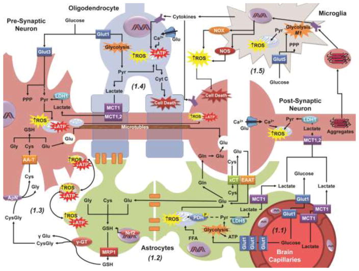
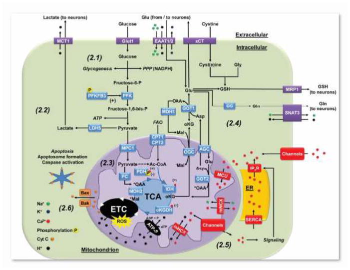
Mitochondrial dysfunction is central to the pathogenesis of neurological disorders. Neurons rely on oxidative phosphorylation to meet their energy requirements and thus alterations in mitochondrial function are linked to energy failure and neuronal cell death. Furthermore, in neurons, dysfunctional mitochondria are reported to increase the steady-state levels of reactive oxygen species derived from the leakage of electrons from the electron transport chain. Research aimed at understanding mitochondrial dysfunction and its role in neurological disorders has been primarily geared towards neurons. In contrast, the effects of mitochondrial dysfunction in glial cells' function and its implication for neuronal homeostasis and brain function has been largely understudied. Unlike neurons and oligodendrocytes, astrocytes and microglia do not degenerate upon the impairment of mitochondrial function, as they rely primarily on glycolysis to produce energy and have a higher antioxidant capacity than neurons. However, recent evidence highlights the role of mitochondrial metabolism and signaling in glial cell function. In this work, we review the functional role of mitochondria in glial cells and the evidence regarding its potential role regulating neuronal homeostasis and disease progression.

Keywords: Astrocytes; Calcium; Free fatty acid oxidation; Glycolysis; Inflammation; Microglia; Mitochondria; Oligodendrocytes; Redox.


Figures

Figure 1
Figure 1
Neuronal metabolism, redox homeostasis and signaling are supported by neighboring glial cells. 1.1: Glucose and lactate enter the brain through Glut1 (glucose transporter 1) and MCT1 (monocarboxylate transporter 1) transporters in the vascular epithelium. Glucose (Glut3) and lactate (MCT1 or 2) are uptaken from the extracellular space by neuronal calls to fuel the TCA cycle for the generation of ATP and biosynthesis of essential molecules. 1.2: As a component of the blood brain barrier (BBB), astrocytes uptake glucose from the capillary epithelium via Glut1 as well, converting the majority of pyruvate (Pyr) generated into lactate which is exported by MCT1. Astrocytes also uptake the neurotransmitter glutamate (Glu) from the synaptic cleft via EAAT (excitatory amino acid transporters) to be (a) converted into glutamine (Gln), (b) exchanged for extracellular cystine (Cys) by xCT, (c) feed into the TCA cycle, or (d) for GSH synthesis. Astrocytes form extended networks with other glia (oligodendrocytes and astrocytes) via gap junctions, sharing nutrients and molecular components with cells more distal to the capillaries. 1.3: Astrocytes contribute to the redox state of neuronal cells by exporting GSH via MRP1 which is broken down by γGT and ApN into its amino acid components to be uptaken and reassembled as GSH in neuronal cells. Dysfunctional or damaged mitochondrial, likely capable of generating ROS, are transferred from neurons to astrocytes to be degraded by mitophagy. 1.4: Oligodendrocytes wrap neuronal projections (myelin sheaths) improving signal conduction and like astrocytes, have been proposed to shuttle lactate to the neurons. 1.5: Microglia are activated by a variety of factors, including cytokines, oxidized proteins, and protein aggregates. Activated microglia migrate to the site of damage and can induce neuronal or oligodendrocyte cell death through the release of cytokines, and the generation of ROS via NADPH oxidases (NOX) and nitric oxide synthases (NOS). AA-T, amino acid transporters; LDH1 or 5, lactate dehydrogenase isoform 1 or 5.
Figure 2
Figure 2
Mitochondrial metabolism and signaling in astrocytes. 2.1: Glucose in astrocytes is used for glycogenesis, NADPH production through the PPP, or glycolysis. Astrocytes are highly glycolytic due to the expression of high levels of 6-phosphofructo-2-kinase/fructose-2,6-bisphosphatase-3 (PFKFB3), whose byproduct fructose-2,6-bisphosphate (F2,6P2), is a positive effector of the glycolytic enzyme 6-phosphofructo-1-kinase (PFK1). In addition, the activity of PFKFB3 is increased by phosphorylation by 5′-AMP-activated protein kinase (AMPK) (Bolanos 2016). 2.2: Astrocytes primarily derive ATP from glycolysis rather than oxidative phosphorylation, where pyruvate is converted to lactate by LDH5 and exported to the extracellular space to be consumed by neurons. 2.3: Astrocytes carboxylate pyruvate to oxaloacetate (OAA) via pyruvate carboxylase (PC) to regenerate TCA cycle intermediates. Phosphorylation of pyruvate dehydrogenase (PDH) restricts the conversion of pyruvate to acetyl-CoA (Ac-CoA). Thus, FAO has been proposed to be the primary contributor of Ac-CoA to the TCA cycle. 2.4: Alpha ketoglutarate (αKG) generated from the TCA cycle can be transported to the cytosol and converted to Glu by glutamic-oxaloacetic transaminase 1 or aspartate (Asp) aminotransferase (GOT1) as part of the malate-Asp shuttle. Glu has three central metabolic pathways in astrocytes. 1) Glu can be converted to Gln by GS and exported to neurons by the sodium-coupled neutral amino acid transporter 3 (SNAT3). 2) Glu is exchanged via xCT for extracellular cystine that is reduced to Cys. Extracellular Glu can be uptaken back by astrocytes by EAAT1/2. Finally, 3) Glu, Gly and Cys are precursors of GSH, which is also exported to neurons via MRP1. 2.5: The ER acts as a store for intracellular calcium, where the sarco/endoplasmic reticulum calcium ion ATPase (SERCA) pumps cytosolic Ca2+ into the ER. Ca2+ signaling is tightly regulated by the activation of IP3R that release Ca2+ from ER stores, as well as by the activation of plasma membrane Ca2+ channels. Mitochondria can buffer Ca2+ by its transport across the inner mitochondrial membrane to the matrix viaMCU), while the export is performed by mNCX and mHCX. Mitochondria can also transport Ca2+ in and out of the mitochondria via the activation of distinct Ca2+ permeable channels. In the matrix, Ca2+ stimulates TCA carbon flux by binding to PDH, IDH, and αKGDH, increasing the activity of the ETC and ATP production. 2.6: Cyt C is held close to the inner mitochondrial membrane by cardiolipin (not shown), acting as a component of ETC. Dissociation of Cyt C from cardiolipin, through oxidative or enzymatic means, coupled with permeabilization of the outer mitochondrial membrane by the formation of Bax/Bak oligomeric channels, allows Cyt C to escape into the cytosol. Cytosolic Cyt C associates with apoptotic protease-activating factor 1 (APAF1), forming the apoptosome and leading to the activation of caspases to initiate apoptosis. AGC, aspartate-glutamate carrier; CPT1 or 2, carnitine palmitoyltransferase isoform 1 or 2; MDH1 or 2, malate dehydrogenase isoform 1 or 2; MPC1, mitochondrial pyruvate carrier 1; OGC, 2-oxoglutarate (α-ketoglutarate) carrier.

References

    1. Amaral AI, Hadera MG, Tavares JM, Kotter MR, Sonnewald U. Characterization of glucose-related metabolic pathways in differentiated rat oligodendrocyte lineage cells. Glia. 2016;64:21–34. - PMC - PubMed
    1. Aoyama K, Watabe M, Nakaki T. Regulation of neuronal glutathione synthesis. J Pharmacol Sci. 2008;108:227–238. - PubMed
    1. Ballinger S, Baumann N, Pham-Dinh D Mitochondrial Toxicity of Tobacco Smoke Air Pollution. This Special Issue. Biology of oligodendrocyte and myelin in the mammalian central nervous system. Physiol Rev. 2001;81:871–927. - PubMed
    1. Belanger M, Allaman I, Magistretti PJ. Brain energy metabolism: focus on astrocyte-neuron metabolic cooperation. Cell Metab. 2011;14:724–738. - PubMed
    1. Boitier E, Rea R, Duchen MR. Mitochondria exert a negative feedback on the propagation of intracellular Ca2+ waves in rat cortical astrocytes. J Cell Biol. 1999;145:795–808. - PMC - PubMed

Publication types

MeSH terms

LinkOut - more resources