. 2017 Jun 27;7(1):4275.
doi: 10.1038/s41598-017-04494-y.

Peptides derived from MARCKS block coagulation complex assembly on phosphatidylserine

Affiliations

Peptides derived from MARCKS block coagulation complex assembly on phosphatidylserine

Noah Kastelowitz et al. Sci Rep. .

Abstract

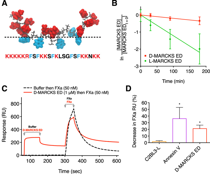
Blood coagulation involves activation of platelets and coagulation factors. At the interface of these two processes resides the lipid phosphatidylserine. Activated platelets expose phosphatidylserine on their outer membrane leaflet and activated clotting factors assemble into enzymatically active complexes on the exposed lipid, ultimately leading to the formation of fibrin. Here, we describe how small peptide and peptidomimetic probes derived from the lipid binding domain of the protein myristoylated alanine-rich C-kinase substrate (MARCKS) bind to phosphatidylserine exposed on activated platelets and thereby inhibit fibrin formation. The MARCKS peptides antagonize the binding of factor Xa to phosphatidylserine and inhibit the enzymatic activity of prothrombinase. In whole blood under flow, the MARCKS peptides colocalize with, and inhibit fibrin cross-linking, of adherent platelets. In vivo, we find that the MARCKS peptides circulate to remote injuries and bind to activated platelets in the inner core of developing thrombi.


Conflict of interest statement

H.Y., R.T., and N.K. have filed patent applications related to this work.

Figures

Figure 1
Figure 1
D-MARCKS ED is a protease resistant peptide that antagonizes the binding of FXa to PS. (A) Sequence and representative membrane bound conformation of the MARCKS ED peptide. Positively charged residues are shown in red and phenylalanine residues are shown in blue. Dashed line shows approximate location of the membrane lipid head groups. (B) Comparison of the human serum stability of L-MARCKS ED (t 1/2 = 57 min, 95% confidence interval (CI) 46 to 79 min) and D-MARCKS ED (t 1/2 = 356 min, 95% CI 252 to 606 min) peptides (n = 3, mean ± SD). (C) Representative reference corrected Biacore 3000 SPR sensogram showing D-MARCKS ED antagonizes the binding of FXa to lipid membrane surface containing PS. Dashed black line shows an injection of running buffer at t = 30 sec and an injection of 50 nM FXa at t = 300 sec. Following complete dissociation of FXa from the membrane surface, the solid red line shows an injection of 1 μM D-MARCKS ED at t = 30 sec and an injection of 50 nM FXa at t = 300 sec. (D) Quantification of FXa SPR binding inhibition with BiOptix 404pi instrument by negative control C2BL3-L, positive control annexin V, and D-MARCKS ED (n = 3 C2BL3-L and annexin V; n = 5 D-MARCKS ED, mean ± SD). *P < 0.05 compared to C2BL3-L by Kruskal-Wallis test followed by Dunn’s post hoc test.
Figure 2
Figure 2
MARCKS ED inhibits prothrombinase enzymatic activity in the presence of PS containing membranes and only binds to activated platelets. (A) Prothrombinase activity of washed human platelets stimulated with thrombin and convulxin when pre-treated with vehicle control or 1 μM L-MARCKS ED, D-MARCKS ED, positive control annexin V, negative control C2BL3-L, or negative control L-MARCKS ED FA mutant (n = 8, mean ± SD). (B) Dose response of D-MARCKS ED pre-treatment on the prothrombinase activity of washed human platelets stimulated with convulxin and thrombin (n = 4, mean ± SD). (C) Prothrombinase activity of exosomes isolated from MDA-MB-231 cells when pre-treated with vehicle control or 1 μM L-MARCKS ED, D-MARCKS ED, annexin V, or C2BL3-L (n = 8, mean ± SD). (D) Prothrombinase activity of synthetic liposomes composed of POPC/POPS at a 19/1 ratio when pre-treated with vehicle control or 1 μM L-MARCKS ED, D-MARCKS ED, annexin V, or C2BL3-L (n = 8, mean ± SD). (E) The binding of annexin V (Brilliant Violet 605) and D-MARCKS ED (NBD) to P-selectin (Cy5) positive platelets was compared by flow cytometry when the platelets were left unstimulated or stimulated with thrombin, convulxin, or thrombin and convulxin (n = 6, mean ± SD). *P < 0.05, **P < 0.01, ***P < 0.001 either by comparison to vehicle control by ANOVA followed by Dunnet’s post hoc test (A,C,D) or two tailed Student’s t-test (E).
Figure 3
Figure 3
MARCKS ED inhibits fibrin formation in whole blood under physiologic flow conditions. (A) Representative images of platelet accumulation (blue, anti-CD41), fibrin(ogen) formation (red, Alexa Flour 647-fibrinogen), and peptide binding (green, NBD-peptide) in the whole blood microfluidic flow assay after 10 min at wall shear rate 100 s−1 when pre-treated with vehicle control or 1 μM L-MARCKS ED, D-MARCKS ED, negative control C2BL3-L, negative control L-MARCKS ED FA mutant, positive control annexin V, or positive control 15 USP ml−1 heparin (n = 6). Scale bar, 50 μm. (B) Time course of peptide NBD fluorescence intensity, platelet surface area coverage, and fibrin(ogen) intensity for the vehicle control and D-MARCKS ED treatment in the microfluidic flow assay (n = 6, mean ± SD). (C) Final fibrin(ogen) intensity values for each treatment in the microfluidic flow assay (n = 6, mean ± SD). (D) Final platelet surface coverage values for each treatment in the microfluidic flow assay (n = 6, mean ± SD). (E) Representative scanning electron micrographs of finals clots formed in microfluidic flow assay with vehicle control and D-MARCKS ED treatment (n = 3). Scale bar, 10 μm. *P < 0.05 compared to vehicle control by ANOVA followed by Dunnet’s post hoc test (C,D).
Figure 4
Figure 4
D-MARCKS ED binds to thrombi in vivo. (A) Representative fluorescence and bright field images of platelet accumulation (blue, anti-CD41), fibrin formation (red, anti-fibrin), and peptide binding (green, NBD-peptide) in the murine intravital laser-induced microvascular injury model when treated with 5 mg kg−1 negative control C2BL3-L or D-MARCKS ED. Scale bar, 10 μm. (BJ) Platelet area, fibrin area, and peptide NBD integrated fluorescence intensity accumulation following murine intravital microvascular injury when treated with 5 mg kg−1 C2BL3-L or D-MARCKS ED. Time course data is shown as mean ± SEM (B,E,H) and median (C,F,I) values. Peak platelet, fibrin, and NBD values (D,G,J) are shown as mean ± SEM (n = 33 thrombi from 4 mice). ***P < 0.001 when compared by the Mann-Whitney statistical test (D,G,J).

References

    1. Bevers EM, Comfurius P, Zwaal RF. Platelet procoagulant activity: physiological significance and mechanisms of exposure. Blood Rev. 1991;5:146–154. doi: 10.1016/0268-960X(91)90031-7. - DOI - PubMed
    1. Jobe SM, et al. Critical role for the mitochondrial permeability transition pore and cyclophilin D in platelet activation and thrombosis. Blood. 2008;111:1257–1265. doi: 10.1182/blood-2007-05-092684. - DOI - PMC - PubMed
    1. Suzuki J, Umeda M, Sims PJ, Nagata S. Calcium-dependent phospholipid scrambling by TMEM16F. Nature. 2010;468:834–838. doi: 10.1038/nature09583. - DOI - PubMed
    1. Bevers EM, Comfurius P, Zwaal RF. Changes in membrane phospholipid distribution during platelet activation. Biochim. Biophys. Acta. 1983;736:57–66. doi: 10.1016/0005-2736(83)90169-4. - DOI - PubMed
    1. Jones ME, Lentz BR, Dombrose FA, Sandberg H. Comparison of the abilities of synthetic and platelet-derived membranes to enhance thrombin formation. Throm. Res. 1985;39:711–724. doi: 10.1016/0049-3848(85)90255-5. - DOI - PubMed

Publication types

MeSH terms

LinkOut - more resources