. 2017 Jun 27;7(1):4304.
doi: 10.1038/s41598-017-04665-x.

Direct conversion of human fibroblasts to brown adipocytes by small chemical compounds

Affiliations

Direct conversion of human fibroblasts to brown adipocytes by small chemical compounds

Yukimasa Takeda et al. Sci Rep. .

Abstract

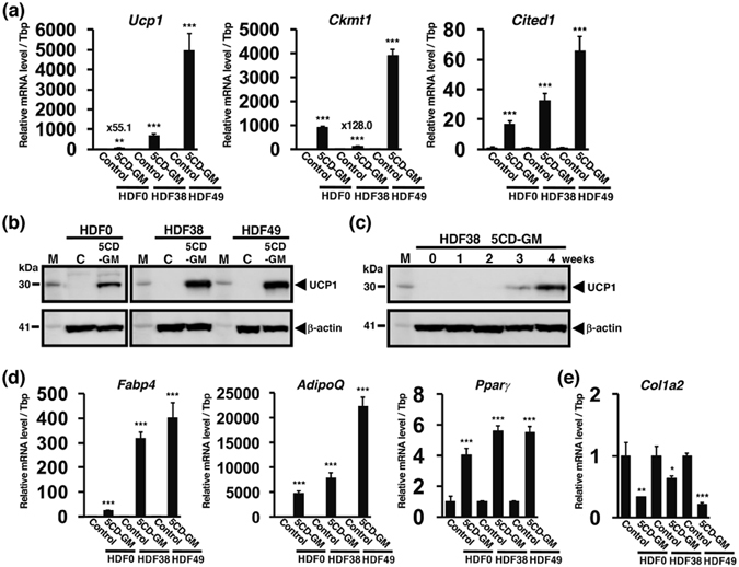
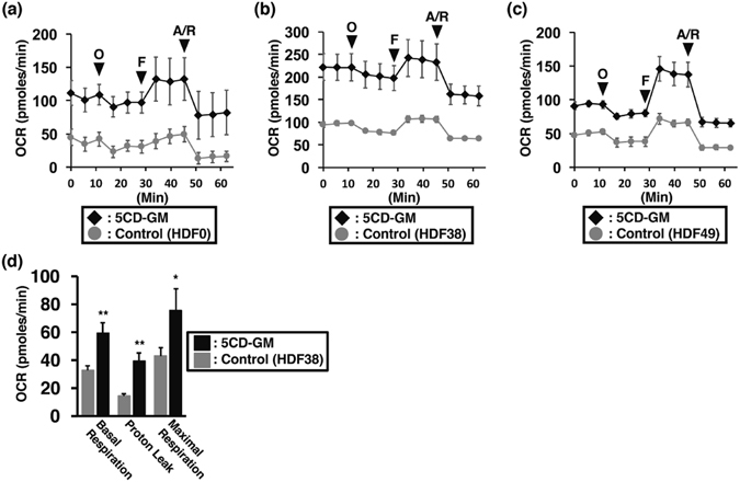
Brown adipocytes play an important role in human energy metabolism and prevention of obesity and diabetes. Induced pluripotent stem cells (iPSCs) represent a promising source for brown adipocytes; however, exogenous gene induction is generally required for iPSCs generation, which might cause undesired effects particularly in long-term treatment after transplantation. We have previously reported a cocktail of six small chemical compounds that enables a conversion of human fibroblasts into chemical compound-induced neuronal cells (CiNCs). Here, we report that modified combinations of the chemical compounds and rosiglitazone, a PPARγ agonist, afforded direct conversion of human fibroblasts into brown adipocytes. The chemical compound-induced brown adipocytes (ciBAs) exhibit induction of human brown adipocyte-specific genes such as Ucp1, Ckmt1, Cited1 and other adipocyte-specific genes such as Fabp4, AdipoQ, and Pparγ. Treatment with either isoproterenol or Forskolin further induced the expression of Ucp1, suggesting that β adrenergic receptor signalling in ciBAs could be functional for induction of thermogenic genes. Moreover, oxygen consumption rates were elevated in ciBAs along with increase of cellular mitochondria. Our findings might provide an easily accessible approach for generating human brown adipocytes from fibroblasts and offer therapeutic potential for the management of obesity, diabetes, and related metabolic disorders.


Conflict of interest statement

The authors declare that they have no competing interests.

Figures

Figure 1
Figure 1
Identification of chemical compounds for direct conversion from HDF38 fibroblasts to adipocyte-like cells. Effects of chemical compounds were evaluated 3 weeks after the treatment. (a) Images after the treatment using the adipocyte medium only or the adipocyte medium supplemented with the 6 chemical compounds (6C). Insets show higher magnification. (b) Morphologies after cultivation with the adipocyte medium and the chemical cocktail excluding either CHIR99021 (G) or CHIR99021 and PD0325901 (GM) from 6 C. Insets show adipocyte-like cells. (c) Images after treatment with the adipocyte medium with 5 CD (5C supplemented with Dorsomorphin (D)) (left panel), and the adipocyte medium with chemical cocktails excluding either G (middle panel) or GM (right panel) from 5CD. Insets show adipocyte-like cells. qRT-PCR analyses of adipocyte-specific genes, Fabp4 (d) and AdipoQ (e), and a brown adipocyte-specific gene, Ucp1 (f), after cultivation with the adipocyte medium only (Control) and the medium supplemented with each combination of the chemical compounds as indicated. (g) UCP1 protein levels were evaluated by western blotting analysis. HDF38 fibroblasts were cultured as described above. β-actin is a loading control. “M” and “C” indicate a molecular weight marker and the control, respectively. Data represent mean ± SD.
Figure 2
Figure 2
Chemically induced brown adipocyte-like cells exhibit a high abundance of lipid droplets and UCP1 expression along with elevated level of cellular mitochondria. The cells were induced by treatment with the adipocyte medium and 5CD-GM for 3 weeks followed by cultivation with the adipocyte medium only for 1 week. (a) Oil Red O staining of the adipocyte-like cells derived from the 3 lines of human fibroblasts, HDF0 (left panel), HDF38 (middle panel), and HDF49 (right panel). Images of bright field, UCP1 protein expression (green), mitochondria labelling with MitoTracker (red), and merged image of UCP1 (green), MitoTracker (red) and DAPI (blue) to visualise nuclei in the fibroblasts of HDF0 (b), HDF38 (c), and HDF49 (d). (e) To evaluate the efficiency of direct conversion by 5CD-GM, the numbers of DAPI-positive cells, adipocyte-like cells with lipid droplets, and UCP1-positive cells were counted in the 3 lines of human fibroblasts, HDF0, HDF38, and HDF49 (n = 3). Data represent mean ± SD.
Figure 3
Figure 3
Human brown adipocyte-specific gene expression in chemical compound-induced brown adipocytes (ciBAs). ciBAs were induced by treatment with the adipocyte medium and 5CD-GM for 3 weeks followed by cultivation with the adipocyte medium only for 1 week. (a) qRT-PCR analyses of human brown adipocyte-specific genes, Ucp1, Ckmt1, and Cited1 in the control cells (the adipocyte medium only) and ciBAs derived from the 3 lines of human fibroblasts, HDF0, HDF38, and HDF49. (b) The protein expression of UCP1 was evaluated by western blotting analysis in ciBAs derived from the 3 lines of human fibroblasts. (c) UCP1 protein levels in HDF38 fibroblasts were evaluated every 1 week after treatment with 5CD-GM. The cells during the period from 3 to 4 weeks were cultured with the adipocyte medium only for the maturation. β-actin is a loading control. “M” and “C” indicate a molecular weight marker and the control, respectively. (d) qRT-PCR analyses of Fabp4, AdipoQ, and Pparγ that are preferentially expressed in adipocytes. (e) qRT-PCR assay of a fibroblast-specific gene Col1a2. Data represent mean ± SD (n = 3). Student’s t-test: *P < 0.05, **P < 0.01, ***P < 0.001.
Figure 4
Figure 4
Induced expression of Ucp1 mRNA by isoproterenol and Forskolin in ciBAs. qRT-PCR analyses of Ucp1 mRNA in ciBAs converted from HDF38 fibroblasts after treatment with either isoproterenol (a) (0.1, 1, and 10 μM) or Forskolin (b) (0.1, 1, and 10 μM) for 3 hrs and 6 hrs as indicated. The expression level of ciBAs treated with dimethyl sulfoxide (DMSO) was used to normalize the expression levels. “C” at the first lane represents the expression level of HDF38 fibroblasts treated with the adipocyte medium only. The expression of other brown adipocyte-specific genes, Ckmt1 (c) and Cited1 (d), was quantified in the same manner as described above. Data represent mean ± SD (n = 3). Student’s t-test: *P < 0.05, **P < 0.01, ***P < 0.001.
Figure 5
Figure 5
Increased oxygen consumption rate (OCR) in ciBAs. OCR was measured using the Flux analyzer in the control cells (the adipocyte medium only, grey circles) and ciBAs (black diamonds) derived from the 3 lines of human fibroblasts, HDF0 (a, n = 5), HDF38 (b, n = 3), and HDF49 (c, n = 4). Oligomycin (O), FCCP (F), and Antimycin A/Rotenone (A/R) were added at indicated times to final concentrations at 2, 0.25, and 0.5 μM, respectively. (d) The OCR for basal respiration, proton leak, and maximal respiration was compared between the control and ciBAs derived from HDF38 fibroblasts (n = 3). Data represent mean ± SD. Student’s t-test: *P < 0.05, **P < 0.01.
Figure 6
Figure 6
A schematic model for a possible connection between signalling pathways regulated by each chemical compound and the direct conversion into ciBAs from human dermal fibroblasts. Our results obtained using SB-431542, an inhibitor of TGF-β signalling pathway, suggest that TGF-β signalling pathway negatively regulates adipogenesis, and that the removal of SB-431542 from the chemical cocktails strongly suppressed the conversion efficiency. BMP signalling pathway regulated by the inhibitors, LDN-193189 and Dorsomorphin, is likely involved in modulation of the activity of downstream SMAD proteins shared with TGF-β signalling pathway. Cellular cAMP induction by Forskolin is closely associated with the activation of Ucp1 and other thermogenic genes through several kinases. Rosiglitazone plays a critical role in adipogenesis for both white and brown adipocytes through PPARγ activation. The chemical combination of 5CD-GM and rosiglitazone might promote gene expression of adipogenic genes and brown adipocyte-specific genes in a coordinated manner, which might be associated with a stable conversion into ciBAs from human dermal fibroblasts.

Similar articles

Cited by

References

    1. Takahashi K, Yamanaka S. Induction of Pluripotent Stem Cells from Mouse Embryonic and Adult Fibroblast Cultures by Defined Factors. Cell. 2006;126:663–676. doi: 10.1016/j.cell.2006.07.024. - DOI - PubMed
    1. Sebban S, Buganim Y. Nuclear Reprogramming by Defined Factors: Quantity Versus Quality. Trends Cell Biol. 2015;26:65–75. doi: 10.1016/j.tcb.2015.08.006. - DOI - PubMed
    1. Yoshihara, M., Hayashizaki, Y. & Murakawa, Y. Genomic Instability of iPSCs: Challenges Towards Their Clinical Applications. Stem Cell Rev, in press. - PMC - PubMed
    1. Jung D-W, Kim W-H, Williams DR. Reprogram or reboot: small molecule approaches for the production of induced pluripotent stem cells and direct cell reprogramming. ACS Chem Biol. 2014;9:80–95. doi: 10.1021/cb400754f. - DOI - PubMed
    1. Daniel MG, Lemischka IR, Moore K. Converting cell fates: generating hematopoietic stem cells de novo via transcription factor reprogramming. Ann N Y Acad Sci. 2016;1370:24–35. doi: 10.1111/nyas.12989. - DOI - PMC - PubMed

Publication types

MeSH terms