Case Reports
. 2017:2017:3074240.
doi: 10.1155/2017/3074240. Epub 2017 Jun 1.

Pediatric Ovarian Growing Teratoma Syndrome

Affiliations
Case Reports

Pediatric Ovarian Growing Teratoma Syndrome

Rebecca M Rentea et al. Case Rep Surg. 2017.

Abstract

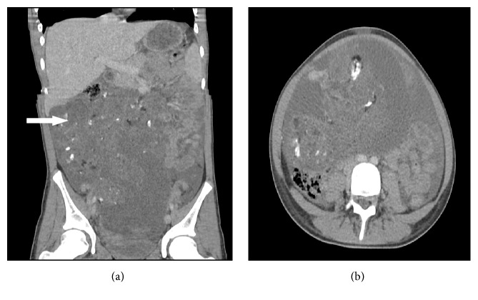
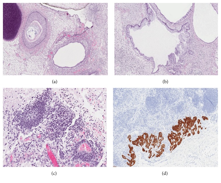
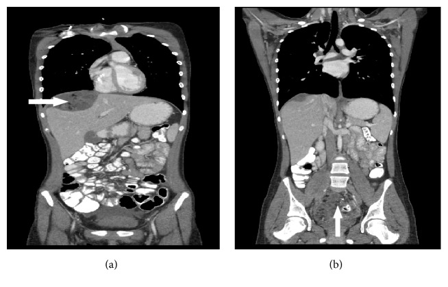
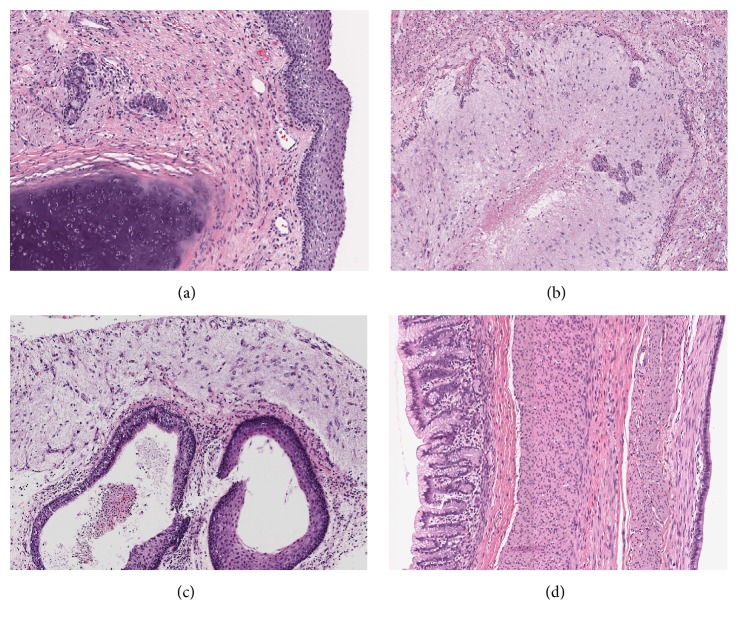
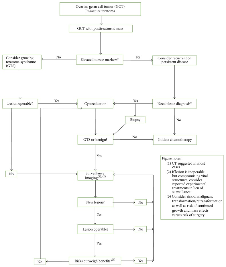
Ovarian immature teratoma is a germ cell tumor that comprises less than 1% of ovarian cancers and is treated with surgical debulking and chemotherapy depending on stage. Growing teratoma syndrome (GTS) is the phenomenon of the growth of mature teratoma elements with normal tumor markers during or following chemotherapy for treatment of a malignant germ cell tumor. These tumors are associated with significant morbidity and mortality due to invasive and compressive growth as well as potential for malignant transformation. Current treatment modality is surgical resection. We discuss a 12-year-old female who presented following resection of a pure ovarian immature teratoma (grade 3, FIGO stage IIIC). Following chemotherapy and resection of a pelvic/liver recurrence demonstrating mature teratoma, she underwent molecular genetics based chemotherapeutic treatment. No standardized management protocol has been established for the treatment of GTS. The effect of chemotherapeutic agents for decreasing the volume of and prevention of expansion is unknown. We review in detail the history, diagnostic algorithm, and previous reported pediatric cases as well as treatment options for pediatric patients with GTS.


Figures

Figure 1
Figure 1
Initial imaging computed tomography of the abdomen and pelvis demonstrating a large pelvic tumor (arrow).
Figure 2
Figure 2
Initial ovarian teratoma with immature elements. (a) Sections of immature teratoma revealed a neoplasm composed of heterogenous elements that included a dermoid cyst, fibroadipose tissue, cartilage, pigmented choroid, and glial tissue (H&E ×100). (b) Cystically dilated glands with enteric differentiation were present (H&E ×100), (c) as well as several foci of immature neuroepithelium (H&E ×200). (d) Immunohistochemical staining with glial fibrillary acid protein (GFAP) reveled teratoma deposits in the periaortic lymph node (GFAP ×200).
Figure 3
Figure 3
Computed tomography abdomen and pelvis follow-up imaging at 8 months. (a) Large mass occupying the right upper abdominal cavity, revealing multiple new masses containing cystic and necrotic elements surrounding the liver (arrow). Because of tumor growth, the giant mass had compressed the liver parenchyma. (b) Pelvic tumor is demonstrated as well (arrow).
Figure 4
Figure 4
Histology of multiple pelvic lesions following repeat laparotomy demonstrated neoplasms composed of derivatives of all three germ layers, namely, ectoderm, mesoderm, and endoderm consistent with mature teratoma. (a) Mature cartilage and squamous epithelium (H&E ×100). (b) Mature neuroglia and vascular proliferation (H&E ×200). (c) Cysts lined with mature squamous and ciliated columnar respiratory epithelium and overlying mature neuroglia (H&E ×200). (d) Large cyst wall with gastric mucinous epithelium, bundles of smooth muscle, and adjacent cysts lining of the ciliated respiratory epithelium (H&E ×200).
Figure 5
Figure 5
Algorithm suggested for management of pediatric female patients with ovarian germ cell tumors following initial resection of immature teratoma and completion of therapy. GCT, germ cell tumor; GTS, growing teratoma syndrome. Modified from Byrd et al. with permission.

Similar articles

Cited by

References

    1. Gorbatiy V., Spiess P., Pisters L. The growing teratoma syndrome: current review of the literature. Indian Journal of Urology. 2009;25(2):186–189. doi: 10.4103/0970-1591.52910. - DOI - PMC - PubMed
    1. Zagamé L., Pautier P., Duvillard P., Castaigne D., Patte C., Lhommé C. Growing teratoma syndrome after ovarian germ cell tumors. Obstetrics and Gynecology. 2006;108(3 I):509–514. doi: 10.1097/01.AOG.0000231686.94924.41. - DOI - PubMed
    1. Lai C.-H., Chang T.-C., Hsueh S., et al. Outcome and prognostic factors in ovarian germ cell malignancies. Gynecologic Oncology. 2005;96(3):784–791. doi: 10.1016/j.ygyno.2004.11.027. - DOI - PubMed
    1. André F., Fizazi K., Culine S., et al. The growing teratoma syndrome: results of therapy and long-term follow-up of 33 patients. European Journal of Cancer. 2000;36(11):1389–1394. doi: 10.1016/S0959-8049(00)00137-4. - DOI - PubMed
    1. Merard R., Ganesan R., Hirschowitz L. Growing teratoma syndrome: A report of 2 cases and review of the literature. International Journal of Gynecological Pathology. 2015;34(5):465–472. doi: 10.1097/PGP.0000000000000180. - DOI - PubMed

Publication types

LinkOut - more resources