. 2017 Jun 21;2(3):e00082-17.
doi: 10.1128/mSphere.00082-17. eCollection 2017 May-Jun.

Sulfide Homeostasis and Nitroxyl Intersect via Formation of Reactive Sulfur Species in Staphylococcus aureus

Affiliations

Sulfide Homeostasis and Nitroxyl Intersect via Formation of Reactive Sulfur Species in Staphylococcus aureus

Hui Peng et al. mSphere. .

Abstract

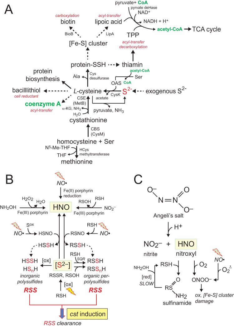
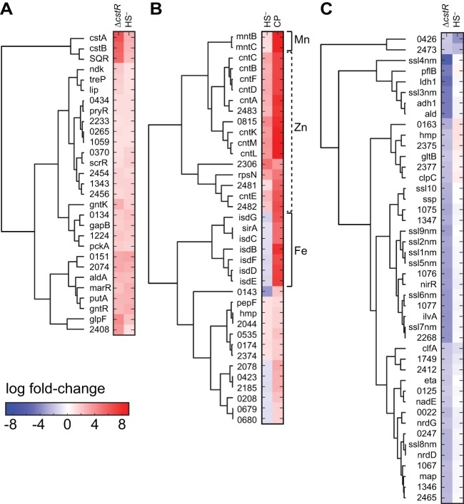
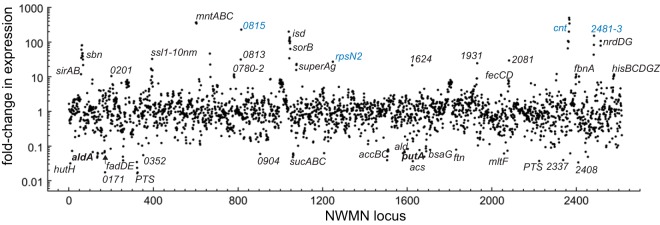
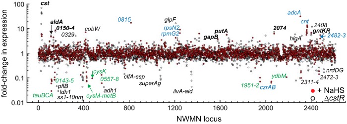
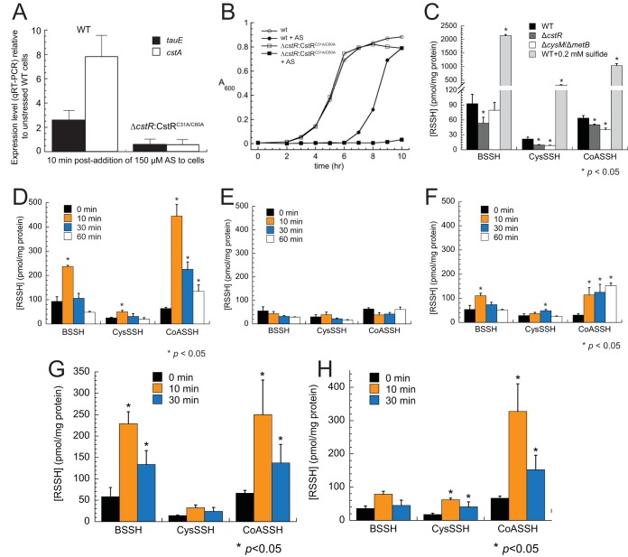
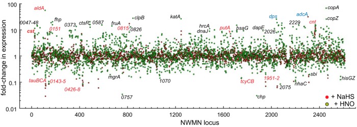
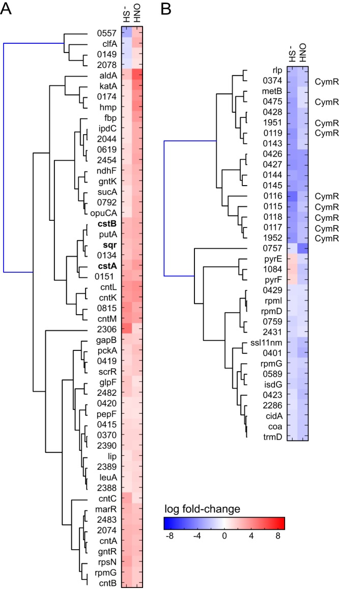
Staphylococcus aureus is a commensal human pathogen and a major cause of nosocomial infections. As gaseous signaling molecules, endogenous hydrogen sulfide (H2S) and nitric oxide (NO·) protect S. aureus from antibiotic stress synergistically, which we propose involves the intermediacy of nitroxyl (HNO). Here, we examine the effect of exogenous sulfide and HNO on the transcriptome and the formation of low-molecular-weight (LMW) thiol persulfides of bacillithiol, cysteine, and coenzyme A as representative of reactive sulfur species (RSS) in wild-type and ΔcstR strains of S. aureus. CstR is a per- and polysulfide sensor that controls the expression of a sulfide oxidation and detoxification system. As anticipated, exogenous sulfide induces the cst operon but also indirectly represses much of the CymR regulon which controls cysteine metabolism. A zinc limitation response is also observed, linking sulfide homeostasis to zinc bioavailability. Cellular RSS levels impact the expression of a number of virulence factors, including the exotoxins, particularly apparent in the ΔcstR strain. HNO, like sulfide, induces the cst operon as well as other genes regulated by exogenous sulfide, a finding that is traced to a direct reaction of CstR with HNO and to an endogenous perturbation in cellular RSS, possibly originating from disassembly of Fe-S clusters. More broadly, HNO induces a transcriptomic response to Fe overload, Cu toxicity, and reactive oxygen species and reactive nitrogen species and shares similarity with the sigB regulon. This work reveals an H2S/NO· interplay in S. aureus that impacts transition metal homeostasis and virulence gene expression. IMPORTANCE Hydrogen sulfide (H2S) is a toxic molecule and a recently described gasotransmitter in vertebrates whose function in bacteria is not well understood. In this work, we describe the transcriptomic response of the major human pathogen Staphylococcus aureus to quantified changes in levels of cellular organic reactive sulfur species, which are effector molecules involved in H2S signaling. We show that nitroxyl (HNO), a recently described signaling intermediate proposed to originate from the interplay of H2S and nitric oxide, also induces changes in cellular sulfur speciation and transition metal homeostasis, thus linking sulfide homeostasis to an adaptive response to antimicrobial reactive nitrogen species.

Keywords: hydrogen sulfide; nitric oxide; nitroxyl; persulfide; reactive nitrogen species; reactive sulfur species; transcriptomics.


Figures

FIG 1
FIG 1
Sulfide (S2−) homeostasis and small-molecule chemistry that couples reactive sulfur species (RSS) and reactive nitrogen species (RNS). (A) Sulfide (S2−, HS, and H2S) and O-acetyl-l-serine (OAS) are substrates for cysteine synthase (CysK) to form l-cysteine. l-Cysteine is the biosynthetic precursor to the major cellular reducing thiol in S. aureus, bacillithiol (BSH), and to coenzyme A (CoASH), which plays important roles in acyl-transfer reactions. l-Cysteine is also the precursor to other major sulfur-containing cofactors, including [Fe-S] clusters, biotin, lipoic acid, and thiamine pyrophosphate (TPP). Lipoic acid and TPP function in the pyruvate dehydrogenase (pyruvate deHase) complex (upper right), used to synthesize acetyl-CoA, which is fed into the tricarboxylic acid (TCA) cycle and other cellular processes. S2– can be accumulate in cells either from exogenous sulfide sources or endogenously via the activity of two enzymes in a transsulfuration pathway, cystathionine β-synthase (CBS) and cystathionine γ-lyase (CSE), which converts homocysteine to l-cysteine. (B) Nitroxyl (HNO) defines an intersection of sulfide, LMW thiols (RSH), reactive sulfur species (RSS), reactive oxygen species (ox), and nitric oxide (NO·). Sulfide can lead to the accumulation of inorganic polysulfides (HSnH) and of organic persulfides (n = 1) and polysulfides (RSSnH), collectively termed reactive sulfur species (RSS), for which there is evidence in mammalian cells (16) and bacterial cells (26). These RSS are sensed by CstR (29), leading to the upregulation of a sulfide oxidation system that includes a canonical sulfide:quinone oxidoreductase (SQR) (26, 81). The RSS can also be derived from exogenous or endogenous NO·, which reacts with oxidized sulfur species (Sox, RSox) to create the nitrosothiol thionitrous acid (HSNO) and organic nitrosothiols (RSNO), which in turn react with S2– to make HNO (82-86). HNO can also be made via 1-electron reduction of NO· or other transformations (top of figure; note that not all possible reactions are shown [85]). (C) Angeli’s salt (AS) is an HNO donor (34) which, at pH values between 4 and 8, decomposes to HNO (pKa, 11.4) (87) and nitrite (NO2). HNO further reacts with O2 to create the potent oxidant peroxynitrite (ONOO), which in turn can react with S2– of [Fe-S] clusters to give perthionitrite (SSNO) (83), thus leading to the decomposition of protein-bound [Fe-S] clusters. SSNO is unstable in aqueous solution and may regenerate HNO (83). HNO is also a highly reactive electrophile at neutral pH that reacts with protein thiols to form sulfinamides in aqueous solution (56); this reaction is in competition with HNO dimerization to form N2O and H2O (66) (not shown).
FIG 2
FIG 2
Clustering analysis and RNAseq transcriptomic analysis of Staphylococcus aureus strain Newman. Genes that change expression significantly in pairwise comparisons of results of sulfide (HS) treatment versus the ΔcstR strain (upregulated genes only) (A) or of sulfide treatment versus CP treatment (all genes) (B) are indicated. (C) A list of genes in the ΔcstR strain (Table 4) that are significantly downregulated compared to their expression under sulfide stress conditions. Genes are clustered according to similarities in changes in expression in each pair of experiments.
FIG 3
FIG 3
RNAseq transcriptomic analysis of wild-type Staphylococcus aureus strain Newman treated with calprotectin (CP). The fold change in expression for each locus tag is indicated (see Table S1A for a complete list of genes induced ≥2.0-fold and with adjusted P values of ≤0.05). Gene names are indicated where known; otherwise, the locus identifier (NWMN_wxyz [where "wxyz" represents the locus number]) is indicated. Names in blue are genes also induced by sulfide (Fig. 4).
FIG 4
FIG 4
RNAseq transcriptomic analysis of Staphylococcus aureus strain Newman. Cells were either treated with 0.2 mM NaHS (red filled circles) or left untreated (ΔcstR strain) (black open symbols); data are expressed relative to the untreated wild-type strain results. The fold change in expression for each locus tag (NWMN_wxyz) is indicated (see Tables 1 to 4 for partial lists of these genes and Table S1A for a complete list). Gene names are indicated where known. Black bold type is used to represent genes that change expression in sulfide-treated cells, in Angeli’s salt (AS; nitroxyl)-treated cells, and in the ΔcstR strain; light blue type is used to represent genes observed to change in the calprotectin-treated samples; green type is used to represent genes of the CymR regulon (3).
FIG 5
FIG 5
Angeli’s salt (AS) induces expression of the cst operon in a CstR-dependent manner and gives rise to a transient increase in endogenous LMW thiol persulfide levels, attributable to HNO. (A and B) AS induces cst operon expression as measured by qRT-PCR (A) and gives rise to a measurable growth phenotype when cells express an inactive CstR, C31A/C60A CstR (29) (B). WT, wild type. (C) LMW persulfide levels can be manipulated by genetic background and exogenous sulfide exposure. (D and E) AS (D) causes increased levels of LMW persulfides under aerobic conditions but nitrite (E) does not, indicating that HNO induces a transient increase in cellular RSS in S. aureus. (F) Levels of LMW persulfides in a ΔcysM ΔmetB strain were also transiently increased by AS. (G and H) Both AS treatment under microaerophilic conditions (G) and ONOO treatment under aerobic conditions (H) cause an increase in the cellular accumulation of LMW persulfides. Error bars represent standard deviations of results of triplicate biological experiments, with statistical significance relative to the results seen with untreated wild-type cells (C) or to the results seen with each of the endogenous LMW persulfides at 0 min (D to H) established using paired t tests (*, P ≤ 0.05). Note that the quantitations of [RSSH] for t = 0 min in panels C, D, E, and H differ slightly from one another, reflecting the culture-to-culture variability of the measurements. Over all 12 replicates, [BSSH] = 77.1 ± 10.5 pmol/mg protein, [CysSSH] = 22.9 ± 2.6 pmol/mg protein, and [CoASSH] = 64.3 ± 3.0 pmol/mg protein, values fully consistent with previously published findings (26).
FIG 6
FIG 6
RNAseq transcriptomic analysis of Staphylococcus aureus strain Newman cells treated with sulfide (red filled circles) versus Angeli’s salt (green filled circles). The fold change in expression for each locus tag is indicated (Tables 1 to 2; Table S1A). Gene names are indicated where known. Expression of those highlighted in red text was significantly induced or repressed under both experimental conditions; expression of those highlighted in light blue was induced during calprotectin (CP) and Angeli’s salt (nitroxyl) treatment.
FIG 7
FIG 7
Clustering analysis of the RNAseq results based on pairwise comparisons of gene expression in sulfide (HS)-treated versus AS (HNO)-treated S. aureus strain Newman cells. (A) Genes that were upregulated by HNO compared to the analogous change in HS-treated cells. (B) Genes that were downregulated by HNO compared to the analogous change in HS-treated cells. Genes of the cst operon are in boldface (panel A), while those genes previously identified as part of the CymR regulon (3) are marked "CymR" (panel B). Genes are clustered according to similarity in change in expression in each pair of experiments. Only the genes that were affected by both sulfide and AS stress are shown here (Tables 1 and 2, Fig. S9, and Table S1A for a compilation of all transcriptomic changes observed under these conditions).

Similar articles

Cited by

References

    1. Wang T, Leyh TS. 2012. Three-stage assembly of the cysteine synthase complex from Escherichia coli. J Biol Chem 287:4360–4367. doi:10.1074/jbc.M111.288423. - DOI - PMC - PubMed
    1. Ayala-Castro C, Saini A, Outten FW. 2008. Fe-S cluster assembly pathways in bacteria. Microbiol Mol Biol Rev 72:110–125, table of contents. doi:10.1128/MMBR.00034-07. - DOI - PMC - PubMed
    1. Soutourina O, Poupel O, Coppée JY, Danchin A, Msadek T, Martin-Verstraete I. 2009. CymR, the master regulator of cysteine metabolism in Staphylococcus aureus, controls host sulphur source utilization and plays a role in biofilm formation. Mol Microbiol 73:194–211. doi:10.1111/j.1365-2958.2009.06760.x. - DOI - PubMed
    1. Tanous C, Soutourina O, Raynal B, Hullo MF, Mervelet P, Gilles AM, Noirot P, Danchin A, England P, Martin-Verstraete I. 2008. The CymR regulator in complex with the enzyme CysK controls cysteine metabolism in Bacillus subtilis. J Biol Chem 283:35551–35560. doi:10.1074/jbc.M805951200. - DOI - PubMed
    1. Ji Q, Zhang L, Sun F, Deng X, Liang H, Bae T, He C. 2012. Staphylococcus aureus CymR is a new thiol-based oxidation-sensing regulator of stress resistance and oxidative response. J Biol Chem 287:21102–21109. doi:10.1074/jbc.M112.359737. - DOI - PMC - PubMed