. 2017 Jun 21;2(3):e00137-17.
doi: 10.1128/mSphere.00137-17. eCollection 2017 May-Jun.

Human Cytomegalovirus-Infected Glioblastoma Cells Display Stem Cell-Like Phenotypes

Affiliations

Human Cytomegalovirus-Infected Glioblastoma Cells Display Stem Cell-Like Phenotypes

Che Liu et al. mSphere. .

Abstract

Glioblastoma multiforme (GBM) is the most common brain tumor in adults. Human cytomegalovirus (HCMV) genomes are present in GBM tumors, yielding hope that antiviral treatments could prove therapeutic and improve the poor prognosis of GBM patients. We discovered that GBM cells infected in vitro with HCMV display properties of cancer stem cells. HCMV-infected GBM cells grow more slowly than mock-infected controls, demonstrate a higher capacity for self-renewal determined by a sphere formation assay, and display resistance to the chemotherapeutic drug temozolomide. Our data suggest that HCMV, while present in only a minority of the cells within a tumor, could contribute to the pathogenesis of GBMs by promoting or prolonging stem cell-like phenotypes, thereby perpetuating tumors in the face of chemotherapy. Importantly, we show that temozolomide sensitivity is restored by the antiviral drug ganciclovir, indicating a potential mechanism underlying the positive effects observed in GBM patients treated with antiviral therapy. IMPORTANCE A role for HCMV in GBMs remains controversial for several reasons. Some studies find HCMV in GBM tumors, while others do not. Few cells within a GBM may harbor HCMV, making it unclear how the virus could be contributing to the tumor phenotype without infecting every cell. Finally, HCMV does not overtly transform cells in vitro. However, tumors induced by other viruses can be treated with antiviral remedies, and initial results indicate that this may be true for anti-HCMV therapies and GBMs. With such a poor prognosis for GBM patients, any potential new intervention deserves exploration. Our work here describes an evidence-based model for how HCMV could contribute to GBM biology while infecting very few cells and without transforming them. It also illuminates why anti-HCMV treatments may be beneficial to GBM patients. Our observations provide blueprints for future in vitro studies examining how HCMV manipulates stem cell-specific pathways and future clinical studies of anti-HCMV measures as GBM therapeutics.

Keywords: brain cancer; cancer; chemoresistance; chemotherapy; herpesvirus.


Figures

FIG 1
FIG 1
Viral genomes are not detected after ex vivo culture of HCMV-positive GBM tumors. (A) DNA isolated from snap-frozen GBM tumors was used as the template for PCR amplification of the HCMV Us28 gene or cellular actin. PCR products were analyzed by agarose gel electrophoresis with ethidium bromide staining. HCMV TB40/E-infected fibroblasts were used as a positive control (+). Mock-infected fibroblasts were used as a negative control. (B) GBM 112 cells were grown in monolayer culture for the indicated number of passages (passage 0 [p0] to passage 3 [p3]) and analyzed as described above. (C) GBM 112, GBM 114, and GBM 120 cells were grown as spheres for the indicated number of passages and analyzed as described above. Images are representative of triplicate PCR experiments.
FIG 2
FIG 2
Primary GBM cells are permissive to HCMV in vitro. (A) GBM 112 and GBM 114 cells were infected with HCMV TB40/E-mCherry at the indicated MOI for 24 h and then analyzed by flow cytometry for mCherry expression (brown histograms). Mock-infected cells (gray histogram) served as a control. The image is representative of three biological replicates. (B) The average percentage of mCherry-positive (HCMV-infected) cells at 1 day (D1) or 7 days (D7) postinfection for three biological replicates of the experiment presented in panel A are presented. Error bars represent the standard errors of the means for the three biological replicates. (C) GBM 112 spheres infected for 5 days at a MOI of 1.0 with HCMV TB40/E-mCherry (red) and counterstained with 4′,6′-diamidino-2-phenylindole (DAPI) (blue) were imaged by confocal fluorescence microscopy. (D) GBM 112 spheres infected for 7 days at a MOI of 1.0 with HCMV TB40/E-mCherry (red) were imaged by conventional fluorescence microscopy. (Left) Bright-field image; (center) dark-field fluorescence image; (right) bright-field fluorescence image.
FIG 3
FIG 3
Primary GBM cells support productive HCMV replication in vitro. (A) RNA isolated from GBM 112 and GBM 114 cells infected at the indicated MOI with HCMV TB40/E-mCherry for 72 h was analyzed by RT-PCR for the indicated genes. Mock-infected cells (M) were used as the control. The image is representative of three biological replicates. (B) Supernatants collected at the indicated time (in hours postinfection [hpi]) from GBM 112 and GBM 114 cells infected with HCMV TB40/E-mCherry at the indicated MOI were subjected to a standard plaque assay. Duplicate biological assays were performed. (C) Progeny virus from cell-free or cell-associated samples of GBM 112 or GBM 114 cells infected at a MOI of 1.0 for 144 h were quantitated by a plaque assay. Thee biological replicate experiments were performed and analyzed by Student’s t test for statistical significance. **, P < 0.01; ***, P < 0.001.
FIG 4
FIG 4
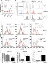
GBM cells infected in vitro with HCMV divide less often than uninfected cells do. (A) GBM 112 cells mock infected or infected at the indicated MOI with HCMV TB40/E-mCherry were grown as spheres, and viable cells were counted at the indicated time (in hours postinfection [hpi]). Averages of three biological replicates are plotted. Values that are significantly different (P < 0.001 by Student’s t test) are indicated (***). (B) Mock-infected or HCMV TB40/E-mCherry-infected GBM cells were loaded with CFSE and then cultured as spheres for 7 days at which time mCherry and CFSE fluorescence of viable cells were quantitated by flow cytometry. For infected cells, the mCherry-negative (mCherry-) and mCherry-positive (mCherry+) populations are shown. The graphs for GBM 112 and GBM 114 cells are representative of three biological replicates. (C) The number of cells within bins representing 1/5th log10 windows is plotted versus the upper limit of the CFSE window for three biological replicates of GBM 112 and GBM 114 cells treated as described above for panel B and one biological replicate of GBM 121 cells. Values that are significantly different by Student’s t test are indicated by asterisks as follows: **, P < 0.01; *, P < 0.05. (D) Cells within the gate representing the top 5% of CFSE staining and mock-infected cells (not shown) were quantified for GBM 112, GBM 114, and GBM 121 cells infected with HCMV at the indicated MOI for 7 days. Averages are plotted with error bars representing the standard errors of the means for biological triplicate samples where appropriate.
FIG 5
FIG 5
GBM cells infected in vitro with HCMV grow as spheres. GBM 112 and GBM 114 cells infected with HCMV TB40/E-mCherry at the indicated MOI were grown as spheres. At the indicated times, images representing randomly selected (presumably different) spheres were captured. Mock-infected cells served as a control.
FIG 6
FIG 6
GBM cells infected in vitro with HCMV show increased sphere-forming ability. (A) GBM 112 and GBM 114 cells infected with HCMV TB40/E-mCherry at the indicated MOI were subjected to a sphere formation assay as described in Materials and Methods. Mock-infected cells were used as the control. The average percentages of sphere-forming cells from three biological replicates are plotted with error bars representing the standard errors of the means. ***, P < 0.001; **, P < 0.01. (B) A serial sphere assay (passage 1 [P1] and passage 2 [P2]) was conducted with GBM 114 cells infected with HCMV TB40/E-mCherry at the indicated MOI, analyzed, and displayed as in panel A. Serially passaged mock-infected cells were used as the control. ***, P < 0.001; NS, not significant by Student’s t test.
FIG 7
FIG 7
GBM 112 and GBM 144 cells infected in vitro with HCMV resist growth inhibition by temozolomide. (A) Sphere formation assays were conducted as described in the legend to Fig. 6 in the presence of carrier (DMSO [D]) or temozolomide (T) where indicated. The average percentages of sphere-forming cells from three biological replicates of  GBM 112 and GBM 144 cells (mock infected or infected with HCMV TB40/E) are plotted with error bars representing the standard errors of the means. ***, P < 0.001; **, P < 0.01; NS, not significant by Student’s t test. (B) Sphere formation assays were conducted as described above with the addition of ganciclovir (G) where indicated. The average percentages of sphere-forming cells from three biological replicates from GBM 112 cells (mock infected or infected with HCMV TB40/E) are plotted with error bars representing the standard errors of the means. ***, P < 0.001; **, P < 0.01; *, P < 0.05; NS, not significant by Student’s t test.

Similar articles

Cited by

References

    1. Ostrom QT, Gittleman H, Fulop J, Liu M, Blanda R, Kromer C, Wolinsky Y, Kruchko C, Barnholtz-Sloan JS. 2015. CBTRUS statistical report: primary brain and central nervous system tumors diagnosed in the United States in 2008–2012. Neuro Oncol 17(Suppl 4):iv1–iv62. - PMC - PubMed
    1. Johnson DR, O’Neill BP. 2012. Glioblastoma survival in the United States before and during the temozolomide era. J Neurooncol 107:359–364. doi:10.1007/s11060-011-0749-4. - DOI - PubMed
    1. Moore PS, Chang Y. 2010. Why do viruses cause cancer? Highlights of the first century of human tumour virology. Nat Rev Cancer 10:878–889. doi:10.1038/nrc2961. - DOI - PMC - PubMed
    1. Chang MH. 2011. Hepatitis B virus and cancer prevention. Recent Results Cancer Res 188:75–84. doi:10.1007/978-3-642-10858-7_6. - DOI - PubMed
    1. Hariri S, Bennett NM, Niccolai LM, Schafer S, Park IU, Bloch KC, Unger ER, Whitney E, Julian P, Scahill MW, Abdullah N, Levine D, Johnson ML, Steinau M, Markowitz LE, HPV-IMPACT Working Group . 2015. Reduction in HPV 16/18-associated high grade cervical lesions following HPV vaccine introduction in the United States − 2008 2012. Vaccine 33:1608–1613. doi:10.1016/j.vaccine.2015.01.084. - DOI - PMC - PubMed

LinkOut - more resources