. 2017 Sep;16(3):2556-2562.
doi: 10.3892/mmr.2017.6858. Epub 2017 Jun 27.

Lentivirus‑mediated RIG‑I knockdown relieves cell proliferation inhibition, cell cycle arrest and apoptosis in ATRA‑induced NB4 cells via the AKT‑FOXO3A signaling pathway in vitro

Affiliations

Lentivirus‑mediated RIG‑I knockdown relieves cell proliferation inhibition, cell cycle arrest and apoptosis in ATRA‑induced NB4 cells via the AKT‑FOXO3A signaling pathway in vitro

Lei Chen et al. Mol Med Rep. 2017 Sep.

Abstract

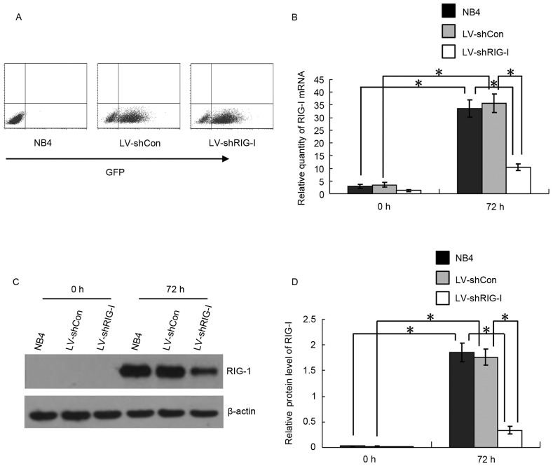
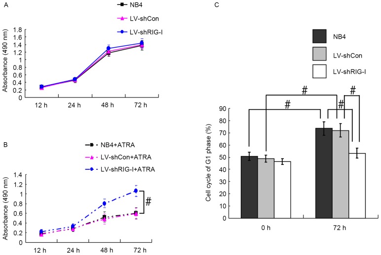
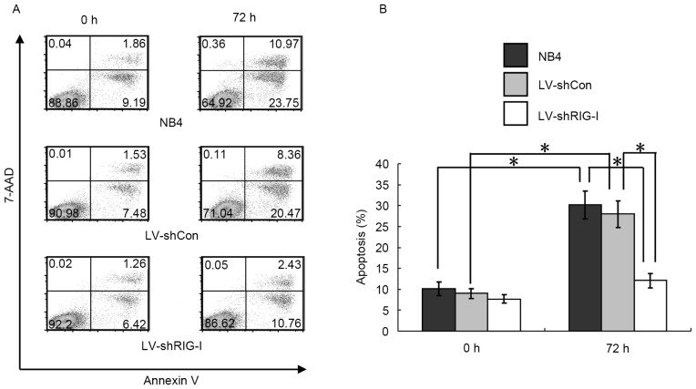
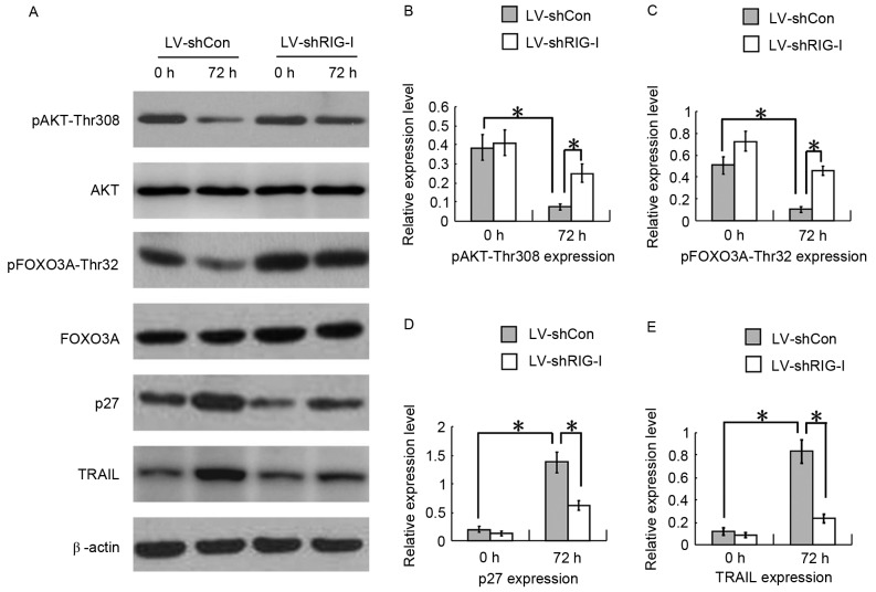
Retinoic acid inducible gene I (RIG‑I) is upregulated during all‑trans retinoic acid (ATRA)‑induced terminal granulocytic differentiation of NB4 acute promyelocytic leukemia (APL) cells. However, the function and mechanism of RIG‑I in NB4 cells remains to be fully elucidated. In the present study, lentivirus‑mediated RIG‑I‑knockdown was used to investigate the proliferation, cell cycle and apoptotic processes of ATRA‑induced NB4 cells in vitro using an MTT assay and flow cytometry, respectively. The roles of RIG‑I and the AKT‑FOXO3A signaling pathway were investigated using western blot analysis. The results showed that the ATRA‑induced expression of RIG‑I was specifically and effectively knocked down at the mRNA and protein levels by lentivirus mediated RIG‑I short hairpin RNA. In addition, silencing of RIG‑I reduced the ATRA‑induced inhibition of NB4 cell proliferation, cell cycle arrest and apoptosis. Further investigations indicated that with ATRA‑induced expression of RIG‑I, levels of phosphorylated (p)AKT‑Thr308 and pForkhead Box (FOX) O3A‑Thr32 were decreased, the expression levels of cell cycle arrest protein p27 and the apoptotic protein, tumor necrosis factor‑related apoptosis‑inducing ligand (TRAIL), directly transcribed by FOXO3A were increased. By contrast, following the knockdown of ATRA‑induced expression of RIG‑I, the levels of pAKT‑Thr308 and pFOXO3A‑Thr32 were increased, and the protein expression levels of p27 and TRAIL were decreased. Taken together, these results showed that the knockdown of RIG‑I reduced the inhibition of cell proliferation, cell cycle arrest and apoptosis in the ATRA‑induced NB4 cells via the AKT‑FOXO3A signaling pathway.


Figures

Figure 1.
Figure 1.
Expression of RIG-I in ATRA-induced NB4 cells is knocked down by LV-mediated shRIG-I. (A) Flow cytometric detection of the expression of GFP in LV-shCon and LV-shRIG-I cells at 72 h post-lentivirus infection. (B) Reverse transcription-quantitative polymerase chain reaction analysis for detection of the RNA expression of RIG-I in NB4, LV-shCon and LV-shRIG-I cells at 0 and 72 h post-ATRA induction (n=3). *P<0.01. (C) Western blot analysis of the protein expression of RIG-I in NB4, LV-shCon and LV-shRIG-I cells at 0 and 72 h post-ATRA induction. (D) Quantitative analysis of protein expression of RIG-I in NB4, LV-shCon and LV-shRIG-I cells at 0 and 72 h post-ATRA induction (n=3) *P<0.01. Data are presented as the mean ± standard deviation. RIG-I, retinoic acid inducible gene I; ATRA, all-trans retinoic acid; GFP, green fluorescent protein; LV, lentivirus; Con, control; siRNA, small interfering RNA; LV-shCon, NB4 cells infected with LV Con-siRNA; LV-shRIG-I, NB4 cells infected with LV RIG-I-siRNA.
Figure 2.
Figure 2.
RIG-I knockdown alleviates ATRA-induced reduced proliferation and cell cycle arrest in NB4 cells. (A) MTT detection of NB4, LV-shCon and LV-shRIG-I cell proliferation without ATRA induction at different time points. (B) MTT detection of NB4, LV-shCon and LV-shRIG-I cell proliferation with ATRA induction at different time points (n=3) #P<0.05. (C) Flow cytometric detection of NB4, LV-shCon and LV-shRIG-I cells in the G1 phase at 0 and 72 h post-ATRA induction (n=3) #P<0.05. Data are presented as the mean ± standard deviation. RIG-I, retinoic acid inducible gene I; ATRA, all-trans retinoic acid; LV, lentivirus; Con, control; siRNA, small interfering RNA; LV-shCon, NB4 cells infected with lentivirus Con-siRNA; LV-shRIG-I, NB4 cells infected with lentivirus RIG-I-siRNA. NB4 + ATRA, ATRA-induced NB4 cells; LV-shCon + ATRA, ATRA-induced LV-shCon cells; LV-shRIG-I + ATRA, ATRA-induced LV-shRIG-I cells.
Figure 3.
Figure 3.
RIG-I knockdown inhibits ATRA-induced apoptosis in NB4 cells. (A) Flow cytometric detection of apoptosis in NB4, LV-shCon and LV-shRIG-I cells at 0 and 72 h post-ATRA induction. (B) Quantitative analysis of apoptosis in NB4, LV-shCon and LV-shRIG-I cells at 0 and 72 h post-ATRA induction (n=3) *P<0.01. Data are presented as the mean ± standard deviation. RIG-I, retinoic acid inducible gene I; ATRA, all-trans retinoic acid; LV, lentivirus; Con, control; siRNA, small interfering RNA; LV-shCon, NB4 cells infected with lentivirus Con-siRNA; LV-shRIG-I, NB4 cells infected with lentivirus RIG-I-siRNA.
Figure 4.
Figure 4.
RIG-I-knockdown reduces cell proliferation inhibition, cell cycle arrest and apoptosis in ATRA-induced NB4 cells via the AKT-FOXO3A signaling pathway. (A) Western blot analyses of expression levels of pAKT-Thr308, pFOXO3A-Thr32, p27 and TRAIL in LV-shCon and LV-shRIG-I cells at 0 and 72 h post-ATRA induction. (B) Quantitative analysis of protein levels of pAKT-Thr308 in LV-shCon and LV-shRIG-I cells at 0 and 72 h post-ATRA induction (n=3) *P<0.01. (C) Quantitative analysis of protein levels of pFOXO3A-Thr32 in LV-shCon and LV-shRIG-I cells at 0 and 72 h post-ATRA induction (n=3) *P<0.01. (D) Quantitative analysis of protein levels of p27 in LV-shCon and LV-shRIG-I cells at 0 and 72 h post-ATRA induction (n=3) *P<0.01. (E) Quantitative analysis of protein levels of TRAIL in LV-shCon and LV-shRIG-I cells at 0 and 72 h post-ATRA induction (n=3) *P<0.01. Data are presented as the mean ± standard deviation. RIG-I, retinoic acid inducible gene I; ATRA, all-trans retinoic acid; LV, lentivirus; Con, control; siRNA, small interfering RNA; LV-shCon, NB4 cells infected with lentivirus Con-siRNA; LV-shRIG-I, NB4 cells infected with lentivirus RIG-I-siRNA.

Similar articles

Cited by

References

    1. Wang ZY, Chen Z. Acute promyelocytic leukemia: From highly fatal to highly curable. Blood. 2008;111:2505–2515. doi: 10.1182/blood-2007-07-102798. - DOI - PubMed
    1. Liu TX, Zhang JW, Tao J, Zhang RB, Zhang QH, Zhao CJ, Tong JH, Lanotte M, Waxman S, Chen SJ, et al. Gene expression networks underlying retinoic acid-induced differentiation of acute promyelocytic leukemia cells. Blood. 2000;96:1496–1504. - PubMed
    1. Zhang NN, Shen SH, Jiang LJ, Zhang W, Zhang HX, Sun YP, Li XY, Huang QH, Ge BX, Chen SJ, et al. RIG-I plays a critical role in negatively regulating granulocytic proliferation. Proc Natl Acad Sci USA. 2008;105:10553–10558. doi: 10.1073/pnas.0804895105. - DOI - PMC - PubMed
    1. Yoneyama M, Kikuchi M, Natsukawa T, Shinobu N, Imaizumi T, Miyagishi M, Taira K, Akira S, Fujita T. The RNA helicase RIG-I has an essential function in double-stranded RNA-induced innate antiviral responses. Nat Immunol. 2004;5:730–737. doi: 10.1038/ni1087. - DOI - PubMed
    1. Zeng W, Sun L, Jiang X, Chen X, Hou F, Adhikari A, Xu M, Chen ZJ. Reconstitution of the RIG-I pathway reveals a signaling role of unanchored polyubiquitin chains in innate immunity. Cell. 2010;141:315–330. doi: 10.1016/j.cell.2010.03.029. - DOI - PMC - PubMed

MeSH terms