. 2017 Aug;38(2):866-874.
doi: 10.3892/or.2017.5761. Epub 2017 Jun 27.

Overexpression of A disintegrin and metalloprotease 10 promotes tumor proliferation, migration and poor prognosis in hypopharyngeal squamous cell carcinoma

Affiliations

Overexpression of A disintegrin and metalloprotease 10 promotes tumor proliferation, migration and poor prognosis in hypopharyngeal squamous cell carcinoma

Chuanjin Ding et al. Oncol Rep. 2017 Aug.

Abstract

The aim of this study was to determine the effect of A disintegrin and metalloprotease 10 (ADAM10) protein expression on the progression, migration and prognosis of hypopharyngeal squamous cell carcinoma (HSCC). Immunohistochemistry and western blot analysis were performed to detect ADAM10 expression in human HSCC specimens. Cell Counting Kit-8 (CCK-8) assay, flow cytometry analysis and wound-healing assay were employed to investigate the effects of ADAM10 knockdown (ADAM10-RNAi) on major oncogenic properties of FaDu cells. We detected that ADAM10 was overexpressed in HSCC specimens and its expression level was associated with differentiation (p<0.001), tumor size (p=0.019), lymph node metastasis (p=0.001), clinical stage (p<0.001), proliferation marker Ki-67 expression (P=0.001) and overall survival (p<0.046). ADAM10-RNAi in FaDu cells resulted in the inhibition of proliferation and the decrease in migration. Moreover, mechanistic experiments revealed that ADAM10-RNAi resulted in an increase in E-cadherin and a decrease in N-cadherin and vimentin expression. Our study implies that high expression of ADAM10 promotes the proliferation and migration of HSCC. These findings may help to provide a method for treatment of HSCC.


Figures
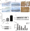
Figure 1.
Figure 1.
Expression level of ADAM10 in paracancerous and HSCC tissues. (A) Representative images of ADAM10 in paracancerous and HSCC tissues. a) Low ADAM10 expression in paracancerous tissue; b) moderate ADAM10 expression in CIS; and c) high ADAM10 expression in IC (magnification ×200, scale bars, 100 µm; magnification ×400, scale bars, 50 µm). (B) The IHC score of ADAM10 expression in samples displayed in A. The mean ± SD was used to express the data. *P<0.05. (C) The protein levels of ADAM10 in 6 out of 15 paired HSCC (T) and non-cancerous hypopharyngeal tissues (N) by western blotting. (D) ADAM10 protein expression was detected abundantly in FaDu cells by western blot analysis. (E) Quantitative results of the western blot analysis in C. *P<0.05. ADAM10, A disintegrin and metalloprotease 10; HSCC, hypopharyngeal squamous cell carcinoma.
Figure 2.
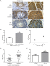
Figure 2.
Relationship between ADAM10 expression and clinicopathological characteristics. (A) Representative images of ADAM10 IHC staining score in different differentiation types, and the relationship between the Ki-67 proliferation index and the ADAM10 expression level (magnification ×200, scale bars, 100 µm; magnification ×400, scale bars, 50 µm). (B) The ADAM10 IHC score was significantly higher in the non-keratinizing type than that in the keratinizing type as shown in A. Moreover, the ADAM10 IHC score was significantly correlated with (C) clinical stages, (D) lymph node metastasis and (E) tumor size. The mean ± SD was used to express the data. *P<0.05. ADAM10, A disintegrin and metalloprotease 10.
Figure 3.
Figure 3.
Scatter plots and Kaplan-Meier overall survival curve. Scatter plots/regression lines in (A) demonstrates a linear correlation for Ki-67 proliferation index and ADAM10 expression in HSCC (Spearmans correlation coefficient P<0.01). (B) Kaplan-Meier overall survival curves for patients with HSCC for low (blue) and high (green) protein expression of ADAM10. Patients in the high expression group had a significantly poorer prognosis than those in the low expression group (P=0.046, log-rank test). ADAM10, A disintegrin and metalloprotease 10; HSCC, hypopharyngeal squamous cell carcinoma.
Figure 4.
Figure 4.
Expression level of ADAM10 in proliferating HSCC FaDu cells. (A) Serum was released into the medium, after cells were serum-starved for 72 h. Flow cytometry was used to detect the percentage of cells in the different phases of the cell cycle. (B) Quantitative results of the percentage of cells in the different phases shown in A. (C) Serum was released into medium, after cells were serum-starved for 72 h. The expression of ADAM10 was analyzed by western blot analysis after cell lysates were collected. (D) Quantitative results of the western blot analysis as shown in C. β-actin was used as a loading control. The mean ± SD was used to express the data. *^#P<0.05 vs. S72 h. ADAM10, A disintegrin and metalloprotease 10; HSCC, hypopharyngeal squamous cell carcinoma.
Figure 5.
Figure 5.
Effects of ADAM10-Si3 on the proliferation of FaDu cells. (A-C) Western blot analysis and real-time PCR were used to detect the effect of ADAM10 knockdown. (D) CCK-8 assay was used to assess cell proliferation. (E and F) Flow cytometry was used to analyze the cell cycle in FaDu cells. Data are expressed as the mean ± SEM. *P<0.05. ADAM10, A disintegrin and metalloprotease 10; CCK-8, Cell Counting Kit-8; ADAM10-Si3, ADAM10-RNA3.
Figure 6.
Figure 6.
Effects of ADAM10-Si3 on the migration of FaDu cells. Migration of FaDu cells was detected by wound-healing assay and Transwell assay after transfection with ADAM10-Si3 or non-specific siRNA or control. (A and B) Decreased healing was observed by wound-healing assay after transfection with ADAM10-Si3. (C and D) Fewer cells passed through the microporous membrane as determined by Transwell assay after transfection with ADAM10-Si3. (E) The expression level of E-cadherin, N-cadherin, and vimentin was detected by western blot analysis in FaDu cells after transfection with ADAM10-Si3. Data are expressed as the mean ± SEM. *P<0.05. ADAM10, A disintegrin and metalloprotease 10; ADAM10-Si3, ADAM10-RNA3.

Similar articles

Cited by

References

    1. Ligier K, Belot A, Launoy G, Velten M, Bossard N, Iwaz J, Righini CA, Delafosse P, Guizard AV. network Francim: Descriptive epidemiology of upper aerodigestive tract cancers in France: Incidence over 1980–2005 and projection to 2010. Oral Oncol. 2011;47:302–307. doi: 10.1016/j.oraloncology.2011.02.013. - DOI - PubMed
    1. Takes RP, Strojan P, Silver CE, Bradley PJ, Jr, Haigentz M, Jr, Wolf GT, Shaha AR, Hartl DM, Olofsson J, Langendijk JA, et al. International Head and Neck Scientific Group: Current trends in initial management of hypopharyngeal cancer: The declining use of open surgery. Head Neck. 2012;34:270–281. doi: 10.1002/hed.21613. - DOI - PubMed
    1. Hoffman HT, Karnell LH, Shah JP, Ariyan S, Brown GS, Fee WE, Glass AG, Goepfert H, Ossoff RH, Fremgen AM. Hypopharyngeal cancer patient care evaluation. Laryngoscope. 1997;107:1005–1017. doi: 10.1097/00005537-199708000-00001. - DOI - PubMed
    1. Godballe C, Jørgensen K, Hansen O, Bastholt L. Hypopharyngeal cancer: Results of treatment based on radiation therapy and salvage surgery. Laryngoscope. 2002;112:834–838. doi: 10.1097/00005537-200205000-00011. - DOI - PubMed
    1. Lee MS, Ho HC, Hsiao SH, Hwang JH, Lee CC, Hung SK. Treatment results and prognostic factors in locally advanced hypopharyngeal cancer. Acta Otolaryngol. 2008;128:103–109. doi: 10.1080/00016480701387116. - DOI - PubMed