. 2017 Jun 28:8:16054.
doi: 10.1038/ncomms16054.

Virus-host relationships of marine single-celled eukaryotes resolved from metatranscriptomics

Affiliations

Virus-host relationships of marine single-celled eukaryotes resolved from metatranscriptomics

Mohammad Moniruzzaman et al. Nat Commun. .

Abstract

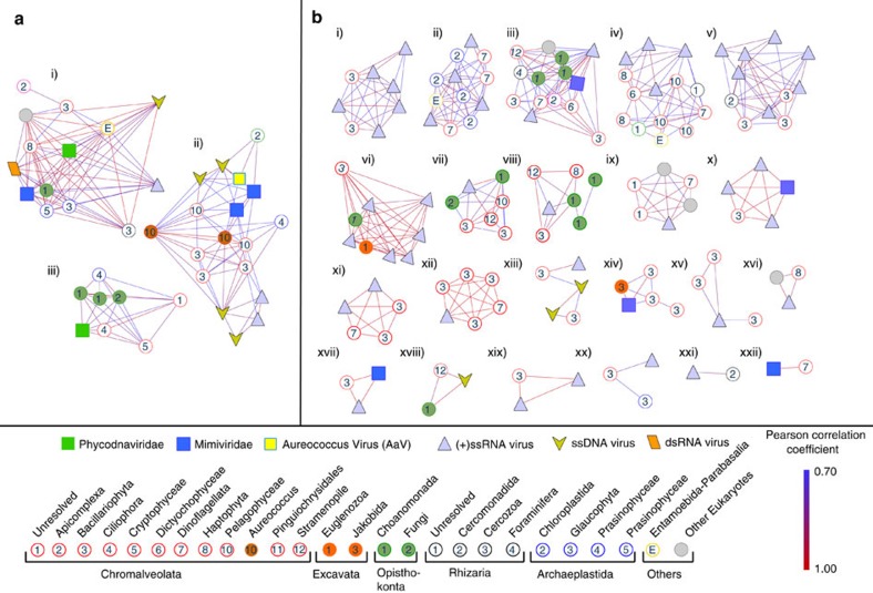
Establishing virus-host relationships has historically relied on culture-dependent approaches. Here we report on the use of marine metatranscriptomics to probe virus-host relationships. Statistical co-occurrence analyses of dsDNA, ssRNA and dsRNA viral markers of polyadenylation-selected RNA sequences from microbial communities dominated by Aureococcus anophagefferens (Quantuck Bay, NY), and diatoms (Narragansett Bay, RI) show active infections by diverse giant viruses (NCLDVs) associated with algal and nonalgal hosts. Ongoing infections of A. anophagefferens by a known Mimiviridae (AaV) occur during bloom peak and decline. Bloom decline is also accompanied by increased activity of viruses other than AaV, including (+) ssRNA viruses. In Narragansett Bay, increased temporal resolution reveals active NCLDVs with both 'boom-and-bust' and 'steady-state infection'-like ecologies that include known as well as novel virus-host interactions. Our approach offers a method for screening active viral infections and develops links between viruses and their potential hosts in situ. Our observations further demonstrate that previously unknown virus-host relationships in marine systems are abundant.


Conflict of interest statement

The authors declare no competing financial interests.

Figures

Figure 1
Figure 1. The abundance of NCLDV core genes within samples.
Core genes from Quantuck Bay (a) and Narragansett Bay (b) are indicated on the X-axes as follows: (A) A32 virion packaging ATPase, (B) VLFT3 like transcription factor, (C) superfamily II helicase II, (D) mRNA capping enzyme, (E) D5 helicase/primase, (F) ribonucleotide reductase small subunit, (G) RNA polymerase large subunit, (H) RNA polymerase small subunit, (I) B-family DNA polymerase and (J) Major Capsid Protein (MCP). Abundance of 9 NCLDV core genes, including MCP, in terms of normalized read counts and number of contigs recovered (up to 100 bp length). The box and whisker plots represent the range of the contig lengths with number of contigs recovered for each gene in brackets. The filled circles represent the rarefied abundances of each core gene in each sample. No contigs could be detected from myristolyated envelope protein, a core NCLDV gene. MCP abundances are shown in red, while other core genes are presented in dark grey.
Figure 2
Figure 2. Phylogenetic reconstruction of NCLDV major capsid protein sequences.
Phylogenetic placement of MCP contigs from (a) Quantuck Bay and (b) Narragansett Bay on a reference tree of NCLDVs with icosahedral capsids. Node support (aLRT-SH statistic) >50% are shown as dark circles. Contigs upto 200 bp are shown, with their expression level RCK in individual samples as a heatmap on the outer rings. Notice that the placement trees contain both AaV reference sequence and the contig originating from AaV (marked with a black arrow). The blue arrow indicates a contig with expression consistent with stable coexistence with the host/persistent infection across multiple time points, while the red arrow denotes a contig with putative ‘boom-and-bust’ like expression. Reference sequences are in bold italic typeface. Abbreviations with NCBI accession numbers: MsV-Marseillevirus (YP_003407071.1), LauV: Lausannevirus (YP_003407071.1), Ws Irido: Weisenia iridescent virus (YP_003407071.1), SG Irido: Singapore Grouper iridescent virus (YP_003407071.1), He Asco: Heliothis virescens Ascovirus (YP_003407071.1), AsfV: African swine fever Virus (NP_042775.1), EhV86: Emiliania huxleyi Virus 86 (NP_042775.1), HaV01: Heterosigma akashiwo Virus 01 (NP_042775.1), PBCV1: Paramacium bursaria Chlorella Virus 1(NP_042775.1), ATCV 1: Acanthocystis turfacea chlorella Virus 1 (NP_042775.1), BpV1: Bathycoccus prasinos Virus 1 (NP_042775.1), MpV12T: Micromonas pusilla Virus 12T (NP_042775.1), OlV1: Ostreococcus lucimarinus Virus 1 (NP_042775.1), AaV: Aureococcus anophagefferens Virus (AaV) (YP_009052173.1), CeV: Chrysochromulina ericina Virus (NP_042775.1), PpV: Phaeocystis pouchetii Virus (NP_042775.1), PgV: Phaeocystis globosa Virus (NP_042775.1), PoV: Pyramimonas orientalis Virus (NP_042775.1), Mega: Megavirus chilensis (NP_042775.1), Moumou: Moumouvirus goulette (AGF85360.1), Mimi: Mimivirus (AAV50707.1), CroV: Cafeteria roenbergensis Virus (YP_003969975.1).
Figure 3
Figure 3. RNA-dependent RNA polymerase phylogenies for sequences attributed to (+) ssRNA viruses.
Phylogenetic placement of (+)ssRNA virus contigs harbouring RNA-dependent RNA polymerase (RdRP) motifs from (a) Quantuck Bay and (b) Narragansett Bay on reference trees. Node support (aLRT-SH statistic) >50% are shown as dark circles. Contigs up to 225 bp are shown, with their expression level (rarefied read counts per kilobase—RCK) in individual samples as a heatmap on the outer rings. The reference sequences are shown in bold italic typeface. Complete name and other details of the reference sequences are presented in Supplementary Data 2.
Figure 4
Figure 4. Complete or near-complete Picornavirales genomes recovered from Quantuck Bay and Narragansett Bay study sites.
Panel (a) shows the phylogenetic classification of these contigs in a topology-only maximum likelihood tree, with contigs from Quantuck Bay having prefix ‘Q_’ and contigs from Narragansett Bay having prefix ‘N_’. Panel (b) shows the genome architecture of these contigs with protein domains and putative CDSs. Panel (c) shows the expression level of these (rarefied read counts per kilobase—RCK) viruses in across different in situ samples.
Figure 5
Figure 5. SIMPROF clustering of virus and eukaryotic markers from Quantuck Bay and Narragansett Bay study sites.
Statistically significant SIMPROF clusters containing both viral and eukaryotic members from (a) Quantuck Bay and (b) Narragansett Bay. Contigs are shown as nodes and the Pearson’s correlations as the connecting edges. Within the SIMPROF clusters, correlations only significant at the level of P≤0.1 are shown. Phylogenetic classifications of the contigs are shown in the bottom panel. A. anophagefferens (dark brown circles) and A. anophagefferens Virus (bright yellow square) are in cluster A(ii).

References

    1. Breitbart M. Marine viruses: truth or dare. Ann. Rev. Mar. Sci. 4, 425–448 (2012). - PubMed
    1. Brum J. R. et al. Patterns and ecological drivers of ocean viral communities. Science 348, 1261498 (2015). - PubMed
    1. Weitz J. S. & Wilhelm S. W. Ocean viruses and their effects on microbial communities and biogeochemical cycles. F1000 Biol. Rep. 4, 17 (2012). - PMC - PubMed
    1. Short S. M. The ecology of viruses that infect eukaryotic algae. Environ. Microbiol. 14, 2253–2271 (2012). - PubMed
    1. Ogata H., Monier A. & Claverie J.-M. in Global Change: Mankind-Marine Environment Interactions eds Ceccaldi H., Dekeyser I., Girault M., Stora G. 157–162Springer Netherlands (2011).

Publication types

LinkOut - more resources