

Fabry disease in donor kidneys with 3- and 12-year follow-up after transplantation
- PMID: 28657066
- PMCID: PMC5477959
- DOI: 10.1093/ndtplus/sfq036
Fabry disease in donor kidneys with 3- and 12-year follow-up after transplantation
Abstract
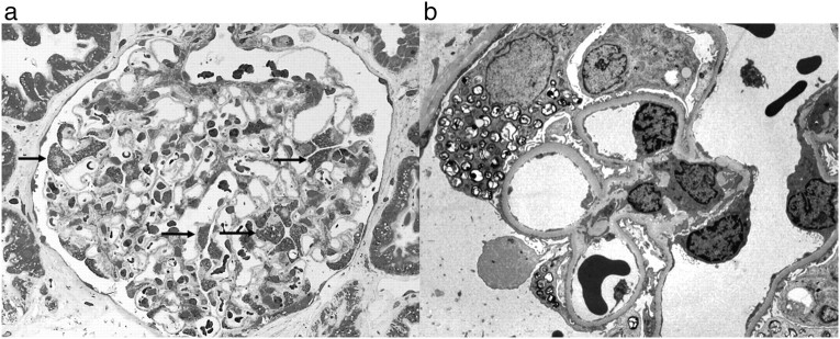
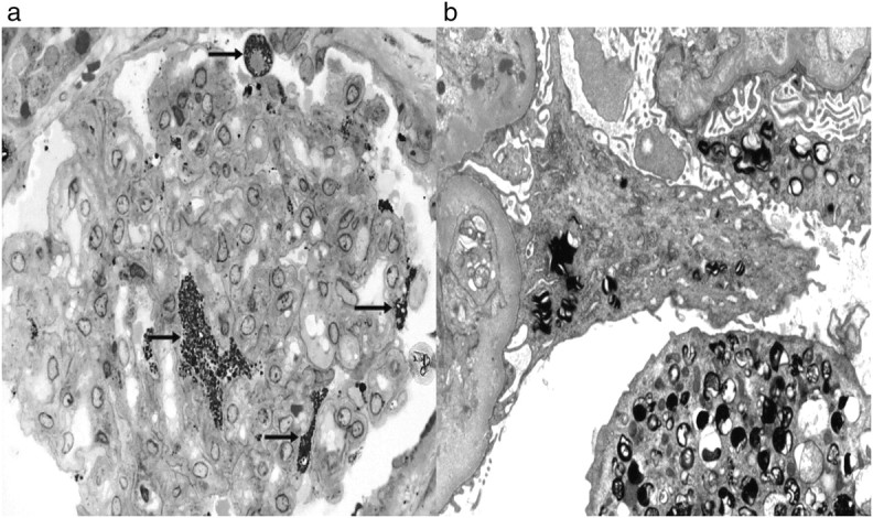
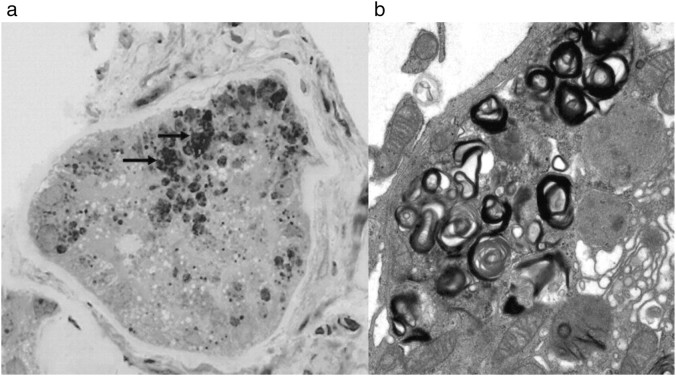
Enzyme replacement therapy (ERT) has been introduced for Fabry disease and has been reported to clear some renal cell types of accumulated glycolipids and to reduce the accumulation in other cell types. We describe two patients without Fabry disease who were transplanted with kidney allografts from a male donor with Fabry disease. Biopsies were taken at transplantation and after 3 years in the first case and after 12 years in the second case. Even though these Fabry kidney allografts for many years had been exposed to normal levels of circulating α-galactosidase A (α-gal-A), the amount of accumulated lysosomal deposits in the podocytes remained unchanged. Additionally, small deposits were also found in tubular cells and glomerular endothelial cells as long as 12 years after transplantation.
Keywords: Fabry disease; enzyme replacement therapy; histological changes; kidney; transplantation.
Figures
References
-
- Sessa A, Meroni M, Battini G, et al. Renal pathological changes in Fabry disease. J Inherit Metab Dis. 2001;24:66–70. - PubMed
-
- Gubler M-C, Lenoir G, Grünfeld J-P, Ulmann A, Droz D, Habib R. Early renal changes in hemizygous and heterozygous patients with Fabry's disease. Kidney Int. 1978;13:223–235. - PubMed
-
- Thurberg BL, Rennke H, Colvin RB, et al. Globotriaosylceramide, accumulation in the Fabry kidney is cleared from multiple cell types after enzyme replacement therapy. Kidney Int. 2002;62:1933–1946. - PubMed
-
- Germain DP, Waldek S, Banikazemi M, et al. Sustained, long-term renal stabilization after 54 months of agalsidase β therapy in patients with Fabry disease. 2007;18:1547–1557. - PubMed
-
- Marshall CB, Shankland S. Cell cycle regulatory proteins in podocyte health and disease. Nephron Exp Nephrol. 2007;106:e51–e59. - PubMed
LinkOut - more resources
Full Text Sources