Review
. 2017 Sep;28(5):e64.
doi: 10.3802/jgo.2017.28.e64. Epub 2017 Jun 5.

Programmed death-1 pathway blockade produces a synergistic antitumor effect: combined application in ovarian cancer

Affiliations
Review

Programmed death-1 pathway blockade produces a synergistic antitumor effect: combined application in ovarian cancer

Xinxin Zhu et al. J Gynecol Oncol. 2017 Sep.

Abstract

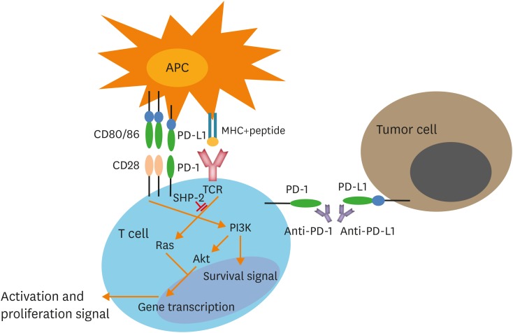
Programmed death-1 (PD-1) and its ligand are part of the immune checkpoint pathway that down-regulates effector T cells in immune response, thereby causing immune suppression. The PD-1/programmed death-ligand 1 (PD-L1) pathway can be blocked by antibodies to reverse tumor-mediated immunosuppression. However, advanced cancers such as stage III-IV ovarian cancer (OC) and certain types such as ID8 OC (a clone of C57BL/6 mouse OC) may hijack the PD-1/PD-L1 pathway to escape immune attack. When combined with chemotherapy, radiotherapy, targeted therapy, immunotherapy, or other agents, these PD-1/PD-L1 pathway blockages can produce a synergistic antitumor response in OC. Combined immunotherapy significantly prolongs overall survival by changing the tumor microenvironment through processes such as increasing the number of CD4⁺ or CD8⁺ T cells or cytokines in mice with OC and decreasing the number of regulatory T cells (Tregs) and myeloid-derived suppressor cells (MDSCs). OC patients treated with combined immunotherapy received better prognoses than those treated with monotherapy. This review reflects the move toward novel therapy combinations for OC and discusses these promising immunotherapeutic approaches, which are more cost-effective and effective than other approaches.

Keywords: Combined Modality Therapy; Immunotherapy; Ovarian Neoplasms; PD-L1 Protein, Human; Programmed Cell Death 1 Receptor; Therapeutic Use; Toxicity.


Conflict of interest statement

No potential conflict of interest relevant to this article was reported.

Figures

Fig. 1
Fig. 1
PD-1 pathway signaling and the targeting of the PD-1 pathway during cancer immunotherapy. T cells recognize the MHC-antigen complex through TCR. CD28 binds to CD80/86 and mediates an activation signal through the PI3K or Ras pathways. PD-1 is ligated with PD-L1 recruitments of SHP-2, which dephosphorylates the downstream molecules and blocks T cell activation. The PD-1 and PD-L1 blockade with mAbs can enhance T cell antitumor activity [28]. APC, antigen-presenting cell; CD, cluster of differentiation; mAbs, monoclonal antibodies; MHC, major histocompatibility complex; PD-1, programmed death-1; PD-L1, programmed death-ligand 1; PI3K, phosphatidylinositol 3-kinase; SHP-2, Src homology region 2 domain-containing phosphatase-2; TCR, T cell receptor.
Fig. 2
Fig. 2
Primary immune checkpoint inhibitors in cells. T cells recognize the MHC-antigen complex through TCR. The ligation of PD-1 with PD-L1/PD-L2 triggers a negative signal in T cells. CTLA-4 competes with CD28 to bind to the costimulatory molecules B7-1 and B7-2 on APCs, mediating an inhibitory signal that leads to the suppression of T cell activation. IgV-like regions are depicted in blue and IgC-like regions in green. APC, antigen-presenting cell; CD, cluster of differentiation; CTLA-4, cytotoxic T-lymphocyte-associated antigen-4; IgC, immunoglobulin constant; IgV, immunoglobulin variable; MHC, major histocompatibility complex; PD-1, programmed death-1; PD-L1, programmed death-ligand 1; PD-L2, programmed death-ligand 2; TCR, T cell receptor.
Fig. 3
Fig. 3
VEGF/VEGFR signaling and the approved therapeutic agents targeting this signaling. VEGF/VEGFR signaling can lead to dysfunctional vasculature that inhibits the infiltration of T cells into the tumor site and reduces DC differentiation and activation. Inhibition of VEGF/VEGFR signaling can improve intratumoral immune cell infiltration and antitumor immune responses [63]. DC, dendritic cell; VEGF, vascular endothelial growth factor; VEGFR, vascular endothelial growth factor receptor.

Similar articles

Cited by

References

    1. Torre LA, Bray F, Siegel RL, Ferlay J, Lortet-Tieulent J, Jemal A. Global cancer statistics, 2012. CA Cancer J Clin. 2015;65:87–108. - PubMed
    1. Siegel R, Naishadham D, Jemal A. Cancer statistics, 2012. CA Cancer J Clin. 2012;62:10–29. - PubMed
    1. Bast RC, Jr, Hennessy B, Mills GB. The biology of ovarian cancer: new opportunities for translation. Nat Rev Cancer. 2009;9:415–428. - PMC - PubMed
    1. Wei H, Zhao L, Hellstrom I, Hellstrom KE, Guo Y. Dual targeting of CD137 co-stimulatory and PD-1 co-inhibitory molecules for ovarian cancer immunotherapy. OncoImmunology. 2014;3:e28248. - PMC - PubMed
    1. Lavoué V, Thédrez A, Levêque J, Foucher F, Henno S, Jauffret V, et al. Immunity of human epithelial ovarian carcinoma: the paradigm of immune suppression in cancer. J Transl Med. 2013;11:147. - PMC - PubMed

MeSH terms

Substances