Review
. 2017 Jun 28;9(7):671.
doi: 10.3390/nu9070671.

Antioxidants Mediate Both Iron Homeostasis and Oxidative Stress

Affiliations
Review

Antioxidants Mediate Both Iron Homeostasis and Oxidative Stress

Mustapha Umar Imam et al. Nutrients. .

Abstract

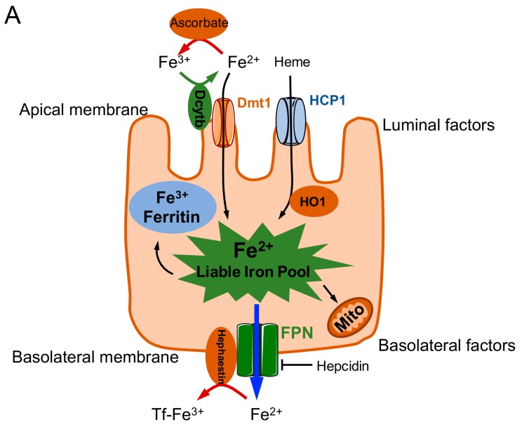
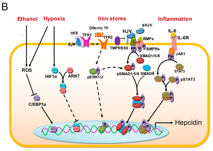
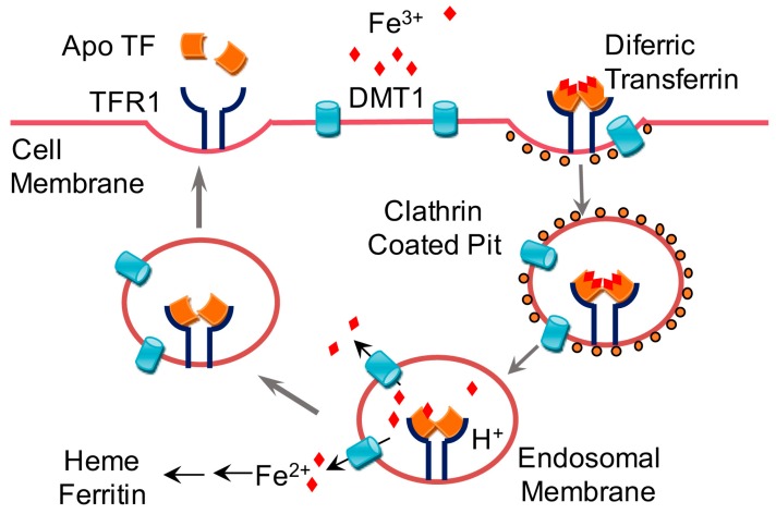
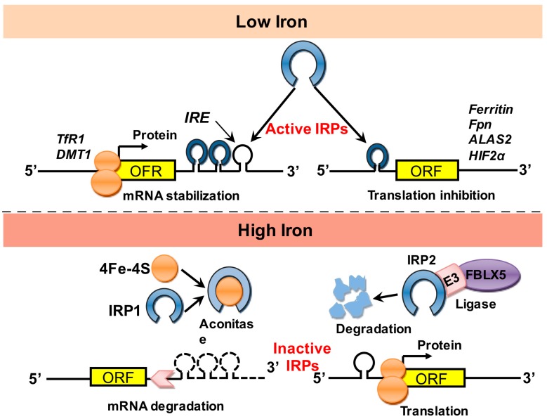
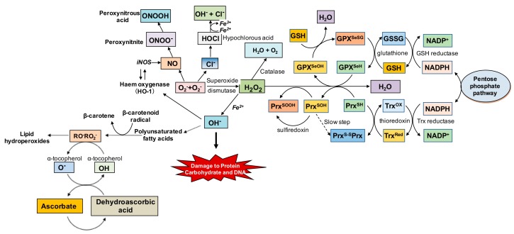
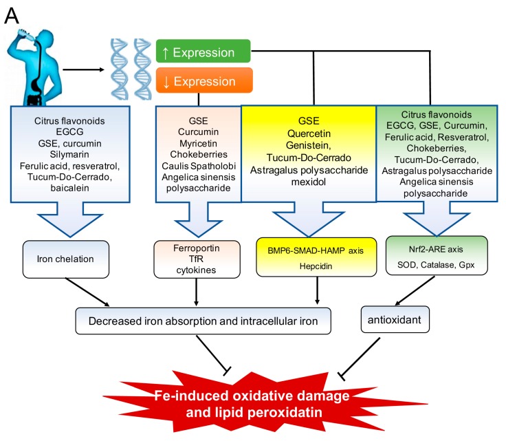
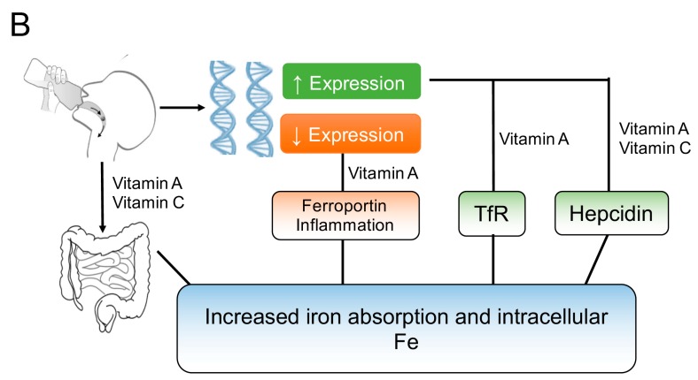
Oxidative stress is a common denominator in the pathogenesis of many chronic diseases. Therefore, antioxidants are often used to protect cells and tissues and reverse oxidative damage. It is well known that iron metabolism underlies the dynamic interplay between oxidative stress and antioxidants in many pathophysiological processes. Both iron deficiency and iron overload can affect redox state, and these conditions can be restored to physiological conditions using iron supplementation and iron chelation, respectively. Similarly, the addition of antioxidants to these treatment regimens has been suggested as a viable therapeutic approach for attenuating tissue damage induced by oxidative stress. Notably, many bioactive plant-derived compounds have been shown to regulate both iron metabolism and redox state, possibly through interactive mechanisms. This review summarizes our current understanding of these mechanisms and discusses compelling preclinical evidence that bioactive plant-derived compounds can be both safe and effective for managing both iron deficiency and iron overload conditions.

Keywords: antioxidants; ferroptosis; iron homeostasis; iron overload; oxidative stress; plant extracts.


Conflict of interest statement

The authors declare no conflicts of interest.

Figures

Figure 1
Figure 1
Schematic overview of iron transport into and across duodenal enterocytes (A) and the pathways involved in regulating transcription of the hepcidin antimicrobial peptide (HAMP) gene to drive hepcidin expression (B). Iron is imported as heme via the heme carrier protein 1 (HCP1) or as Fe2+ (after reduction by duodenal cytochrome b (Dcytb) via the divalent metal transporter (DMT) 1. The labile iron pool within the enterocyte can be stored as ferritin, utilized for mitochondrial oxidative phosphorylation, or exported via ferroportin (Fpn). Hephaestin or ceruloplasmin then converts Fe2+ to Fe3+, which then binds to transferrin (Tf). Hepcidin negatively regulates Fpn. ARNT: aryl hydrocarbon nuclear receptor translocator; β2m:beta-2-microglobulin; BMP: bone morphogenetic protein; BMPR: BMP receptor; C/EBP1α: CCAAT/enhancer-binding protein 1α; ERK1/2: extracellular signal-regulated kinase; HIF1α: hypoxia-inducible factor 1α; HFE: human hemochromatosis protein; HJV: hemojuvelin; HO1: heme oxygenase 1; IL-6: interleukin 6; IL-6R: IL-6 receptor; JAK: Janus kinase; Mito: mitochondria; p: phosphate group; ROS: reactive oxygen species; sHJV: serum HJV; SMAD1/5/8: mothers against decapentaplegic homolog1/5/8; SMAD4: mothers against decapentaplegic homolog 4; STAT3: signal transducer and activator of transcription 3; TfR: Tf receptor; TMPRSS6: transmembrane protease, serine 6.
Figure 1
Figure 1
Schematic overview of iron transport into and across duodenal enterocytes (A) and the pathways involved in regulating transcription of the hepcidin antimicrobial peptide (HAMP) gene to drive hepcidin expression (B). Iron is imported as heme via the heme carrier protein 1 (HCP1) or as Fe2+ (after reduction by duodenal cytochrome b (Dcytb) via the divalent metal transporter (DMT) 1. The labile iron pool within the enterocyte can be stored as ferritin, utilized for mitochondrial oxidative phosphorylation, or exported via ferroportin (Fpn). Hephaestin or ceruloplasmin then converts Fe2+ to Fe3+, which then binds to transferrin (Tf). Hepcidin negatively regulates Fpn. ARNT: aryl hydrocarbon nuclear receptor translocator; β2m:beta-2-microglobulin; BMP: bone morphogenetic protein; BMPR: BMP receptor; C/EBP1α: CCAAT/enhancer-binding protein 1α; ERK1/2: extracellular signal-regulated kinase; HIF1α: hypoxia-inducible factor 1α; HFE: human hemochromatosis protein; HJV: hemojuvelin; HO1: heme oxygenase 1; IL-6: interleukin 6; IL-6R: IL-6 receptor; JAK: Janus kinase; Mito: mitochondria; p: phosphate group; ROS: reactive oxygen species; sHJV: serum HJV; SMAD1/5/8: mothers against decapentaplegic homolog1/5/8; SMAD4: mothers against decapentaplegic homolog 4; STAT3: signal transducer and activator of transcription 3; TfR: Tf receptor; TMPRSS6: transmembrane protease, serine 6.
Figure 2
Figure 2
Schematic depiction of iron trafficking across the plasma membrane into the cell. Transferrin-(Tf-) bound iron is transported into the cell via transferrin receptor 1 (TfR1) or 2 (TfR2). The complex is endocytosed, and a decrease in luminal pH causes the release of iron from the complex. Tf that is completely free of iron (apotransferrin, ApoTf) and the TfR are then recycled back to the cell membrane. The labile pool of iron within the cell exits the endosome via divalent metal transporter 1 (DMT1) and is stored as ferritin. ApoTf: apotransferrin.
Figure 3
Figure 3
Post-transcriptional control of iron homeostasis. Iron-responsive element‒binding proteins (IRP1 and IRP2) bind to the iron-responsive element (IRE) in the untranslated region (UTR) of mRNAs encoding various iron-regulating molecules, thereby regulating their translation. ALAS2: aminolevulinic acid synthase 2; DMT1: divalent metal transporter 1; E3: ubiquitin ligase subunit; FBLX5: F-box and leucine-rich repeat protein 5; Fpn: ferroportin; HIF2α: hypoxia-inducible factor 2α; mRNA: messenger ribonucleic acid; ORF: open reading frame; TfR1: transferrin receptor 1.
Figure 4
Figure 4
Summary of the oxidative stress cascade, including endogenous antioxidant defenses. Superoxide free radicals (O2) generated during metabolic processes are endogenously neutralized by superoxide dismutase to hydrogen peroxide (H2O2), and subsequently to water (H2O) and oxygen (O2), although the glutathione (GSH) system can also neutralize H2O2 to H2O. Conversely, when O2 is not neutralized, it can form more reactive species in the presence of nitric oxide (NO) and chloride (Cl) thereby leading to further oxidative damage. Similarly, H2O2 in the presence of Fe2+ can produce hydroxyl free radicals (OH), which are highly toxic to proteins and DNA, and can even lead to the generation of lipid peroxides that are also prooxidant. DNA: deoxyribonucleic acid; GSSG: glutathione disulfide; NADP: nicotinamide adenine dinucleotide phosphate; NADPH: reduced NADP; RO: alkoxyl radical; RO2: peroxyl radical.
Figure 5
Figure 5
Overview of the mechanisms by which antioxidants regulate iron and oxidative stress. (A) A variety of antioxidants regulate iron absorption by chelating iron and/or by modulating the expression of antioxidant and iron metabolism‒regulating genes, ultimately reducing oxidative damage due to excess iron during iron overload; (B) Vitamins A and C can increase iron absorption and modulate the expression of several proteins (e.g., transferrin receptor (TfR), hepcidin, and ferroportin) and inflammatory signals, providing clinical benefits under iron deficient conditions. BMP6-SMAD-HAMP: bone morphogenetic factor-mothers against decapentaplegic homolog-hepcidin antimicrobial peptide; EGCG: epigallocatechin gallate; GPx: glutathione peroxidase; GSE: grape seed extract; Nrf2-ARE: nuclear factor erythroid 2-related factor 2-antixodant response element; SOD: superoxide dismutase.
Figure 5
Figure 5
Overview of the mechanisms by which antioxidants regulate iron and oxidative stress. (A) A variety of antioxidants regulate iron absorption by chelating iron and/or by modulating the expression of antioxidant and iron metabolism‒regulating genes, ultimately reducing oxidative damage due to excess iron during iron overload; (B) Vitamins A and C can increase iron absorption and modulate the expression of several proteins (e.g., transferrin receptor (TfR), hepcidin, and ferroportin) and inflammatory signals, providing clinical benefits under iron deficient conditions. BMP6-SMAD-HAMP: bone morphogenetic factor-mothers against decapentaplegic homolog-hepcidin antimicrobial peptide; EGCG: epigallocatechin gallate; GPx: glutathione peroxidase; GSE: grape seed extract; Nrf2-ARE: nuclear factor erythroid 2-related factor 2-antixodant response element; SOD: superoxide dismutase.

References

    1. Kohgo Y., Ikuta K., Ohtake T., Torimoto Y., Kato J. Body iron metabolism and pathophysiology of iron overload. Int. J. Hematol. 2008;88:7–15. doi: 10.1007/s12185-008-0120-5. - DOI - PMC - PubMed
    1. Beard J.L., Dawson H., Piñero D.J. Iron metabolism: A comprehensive review. Nutr. Rev. 1998;54:295–317. doi: 10.1111/j.1753-4887.1996.tb03794.x. - DOI - PubMed
    1. Papanikolaou G., Pantopoulos K. Iron metabolism and toxicity. Toxicol. Appl. Pharm. 2005;202:199–211. doi: 10.1016/j.taap.2004.06.021. - DOI - PubMed
    1. Bresgen N., Eckl P.M. Oxidative stress and the homeodynamics of iron metabolism. Biomolecules. 2015;5:808–847. doi: 10.3390/biom5020808. - DOI - PMC - PubMed
    1. Emerit J., Beaumont C., Trivin F. Iron metabolism, free radicals, and oxidative injury. Biomed. Pharmacother. 2001;55:333–339. doi: 10.1016/S0753-3322(01)00068-3. - DOI - PubMed

MeSH terms

LinkOut - more resources