. 2017 Jun 28;9(7):202.
doi: 10.3390/toxins9070202.

Epidermal Growth Factor Receptor Signaling Enhances the Proinflammatory Effects of Staphylococcus aureus Gamma-Toxin on the Mucosa

Affiliations

Epidermal Growth Factor Receptor Signaling Enhances the Proinflammatory Effects of Staphylococcus aureus Gamma-Toxin on the Mucosa

Aaron N Gillman et al. Toxins (Basel). .

Abstract

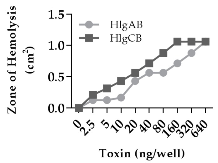
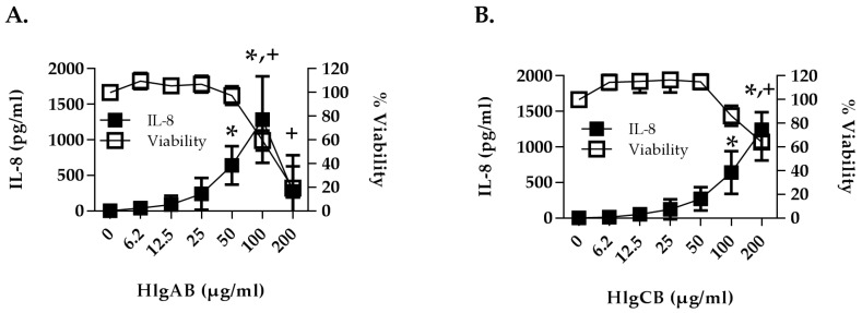
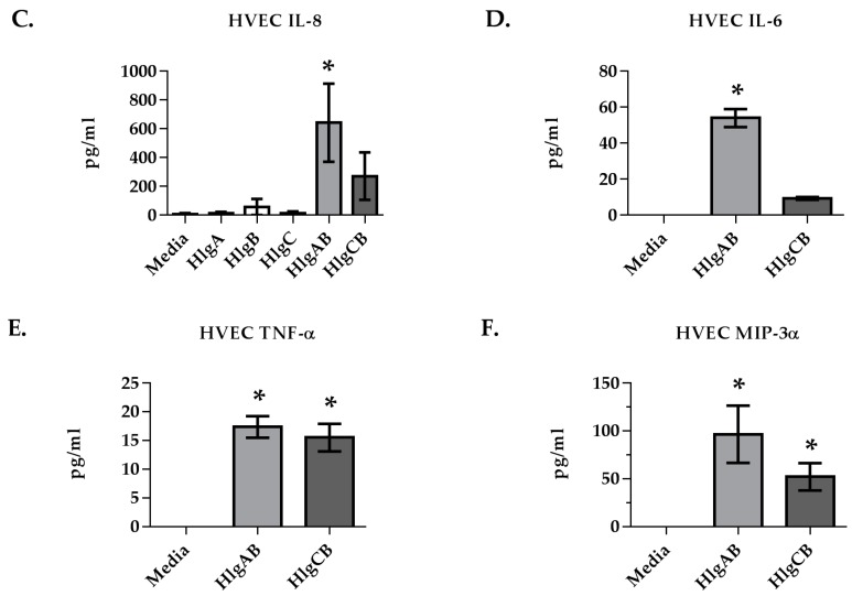
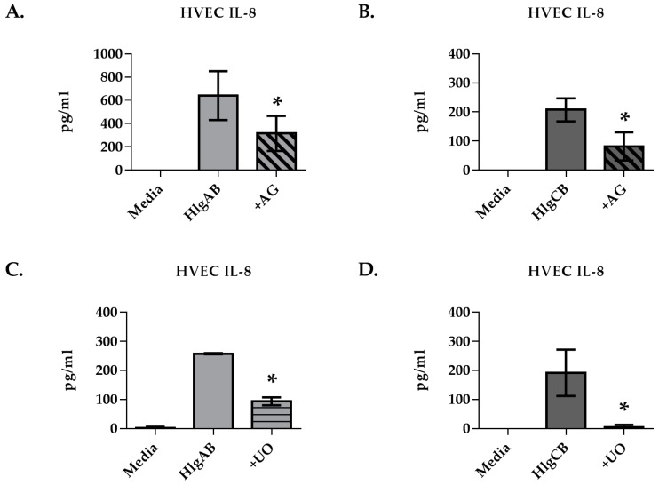
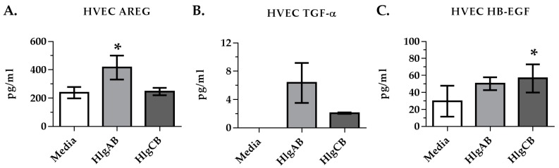
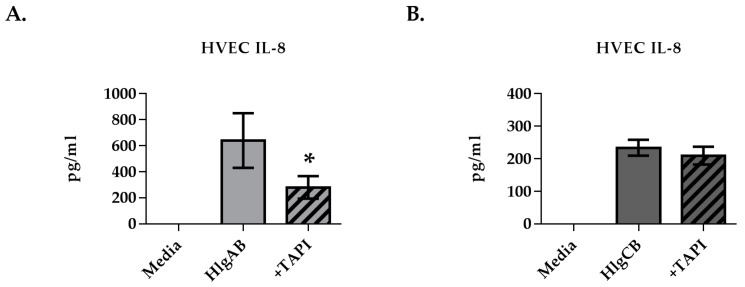
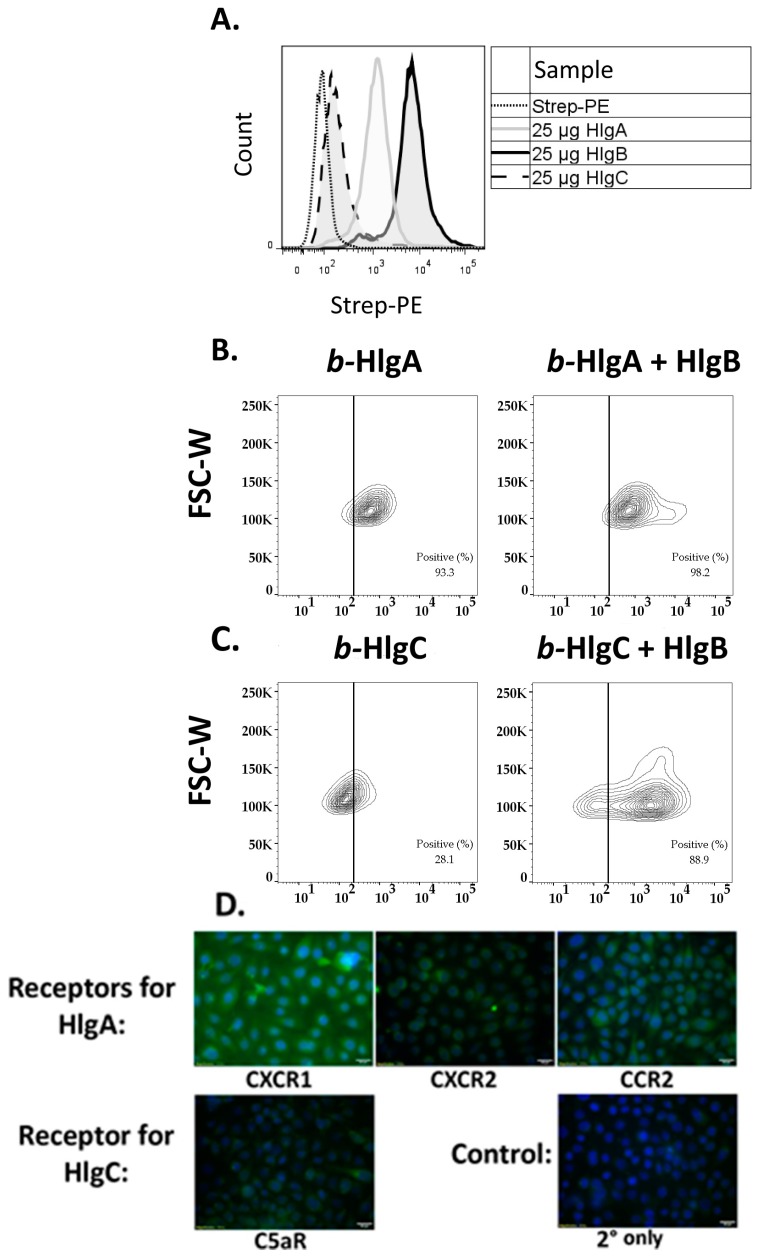
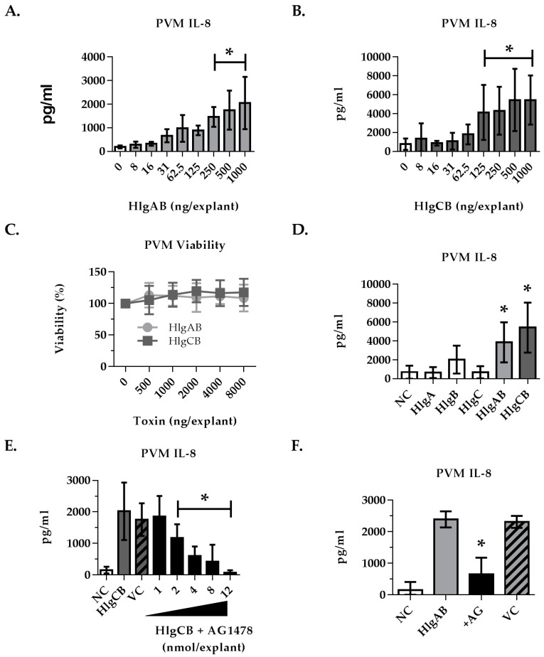
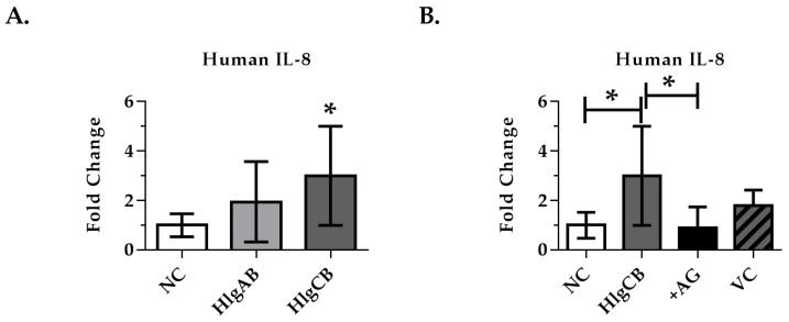
Staphylococcus aureus (S. aureus) produces many different exotoxins including the gamma-toxins, HlgAB and HlgCB. Gamma-toxins form pores in both leukocyte and erythrocyte membranes, resulting in cell lysis. The genes encoding gamma-toxins are present in most strains of S. aureus, and are commonly expressed in clinical isolates recovered from menstrual Toxic Shock Syndrome (mTSS) patients. This study set out to investigate the cytotoxic and proinflammatory effects of gamma-toxins on vaginal epithelial surfaces. We found that both HlgAB and HlgCB were cytotoxic to cultured human vaginal epithelial cells (HVECs) and induced cytokine production at sub-cytotoxic doses. Cytokine production induced by gamma-toxin treatment of HVECs was found to involve epidermal growth factor receptor (EGFR) signaling and mediated by shedding of EGFR ligands from the cell surface. The gamma-toxin subunits displayed differential binding to HVECs (HlgA 93%, HlgB 97% and HlgC 28%) with both components (HlgAB or HlgCB) required for maximum detectable binding and significant stimulation of cytokine production. In studies using full thickness ex vivo porcine vaginal mucosa, HlgAB or HlgCB stimulated a dose-dependent cytokine response, which was reduced significantly by inhibition of EGFR signaling. The effects of gamma-toxins on porcine vaginal tissue and cultured HVECs were validated using ex vivo human ectocervical tissue. Collectively, these studies have identified the EGFR-signaling pathway as a key component in gamma-toxin-induced proinflammatory changes at epithelial surfaces and highlight a potential therapeutic target to diminish toxigenic effects of S. aureus infections.

Keywords: Staphylococcus aureus; and mucosal immune response; epidermal growth factor receptor; gamma-toxin; menstrual toxic shock syndrome; tyrosine kinase inhibitors.


Conflict of interest statement

The authors declare no conflict of interest.

Figures

Figure 1
Figure 1
Gamma-toxin is hemolytic to rabbit erythrocytes. 100 μL of gamma-toxin at a 1:1 molar ratio in Tris-Buffered Saline (TBS) at the indicated dose was placed in 0.3 cm diameter wells within 0.4% agar containing 5% rabbit erythrocytes for 4 h and the zone of hemolysis was measured. Data represent area from individual wells.
Figure 2
Figure 2
Gamma-toxin is cytotoxic and induces production of proinflammatory cytokines in human vaginal epithelial cells (HVECs). HVECs were exposed to (A) HlgAB or (B) HlgCB at the indicated doses for 6 h and processed for cytotoxicity (open squares) by MTT assay or cytokine production (filled squares) by ELISA. For A and B, asterisks (IL-8) and crosses (viability) indicate significant changes (p < 0.05) from media alone controls; (C) IL-8; (D) IL-6; (E) TNF-α and (F) MIP-3α production from HVECs treated with individual subunits of gamma-toxin (50 µg/mL) or HlgAB and HlgCB (50 µg/mL) at a 1:1 molar ratio. Data show mean +/− standard deviation. For D–F, asterisks indicate a significant difference from media alone controls (p < 0.05).
Figure 2
Figure 2
Gamma-toxin is cytotoxic and induces production of proinflammatory cytokines in human vaginal epithelial cells (HVECs). HVECs were exposed to (A) HlgAB or (B) HlgCB at the indicated doses for 6 h and processed for cytotoxicity (open squares) by MTT assay or cytokine production (filled squares) by ELISA. For A and B, asterisks (IL-8) and crosses (viability) indicate significant changes (p < 0.05) from media alone controls; (C) IL-8; (D) IL-6; (E) TNF-α and (F) MIP-3α production from HVECs treated with individual subunits of gamma-toxin (50 µg/mL) or HlgAB and HlgCB (50 µg/mL) at a 1:1 molar ratio. Data show mean +/− standard deviation. For D–F, asterisks indicate a significant difference from media alone controls (p < 0.05).
Figure 3
Figure 3
Epidermal growth factor receptor (EGFR) inhibition reduces significantly gamma-toxin-induced IL-8 production in HVECs. HVECs were exposed to HlgAB (50 µg/mL) or HlgCB (100 µg/mL) at a 1:1 molar ratio for 6 h and IL-8 production was analyzed by ELISA. IL-8 production in response to HlgAB or HlgCB was attenuated in the presence of (A,B) AG1478 or (C,D) UO126. Data show mean +/− standard deviation. Asterisks indicate significant difference from gamma-toxin treated cells with no inhibitor present (p < 0.05).
Figure 4
Figure 4
Gamma-toxin induces shedding of EGFR ligands in HVECs. Cells were incubated with HlgAB (50 µg/mL) or HlgCB (100 µg/mL) at a 1:1 molar ratio for 6 h and processed for shedding of EGFR ligands by ELISA. Data show (A) amphiregulin; (B) tumor growth factor α; and (C) heparin-binding epidermal growth factor mean concentrations +/− standard deviation. Asterisks indicate significant difference from media alone controls (p < 0.05).
Figure 5
Figure 5
ADAM inhibition reduces HlgAB-induced IL-8 production in HVECs. HVECs were exposed to HlgAB (50 µg/mL) or HlgCB (100 µg/mL) at a 1:1 molar ratio for 6 h and processed for IL-8 production by ELISA. TAPI-1 treatment reduced IL-8 production in response to (A) HlgAB but not (B) HlgCB. Data show mean +/− standard deviation. Asterisks indicate significant difference from gamma-toxin-treated cells with no inhibitor present (p < 0.05).
Figure 6
Figure 6
Gamma-toxin HlgB facilitates binding of HlgA and HlgC to HVECs. HVECs were incubated with 25 µg/mL of biotinylated gamma-toxin subunits at 4 °C for 30 min prior to fixation and staining. (A) Relative binding of individual biotinylated subunits on HVECs. Non-biotinylated HlgB significantly increased binding of biotinylated; (B) HlgA or (C) HlgC binding to HVECs (Chi Squared T(X) = 876 – b-HlgC + HlgB and T(X) = 352 for b-HlgA + HlgB). Data show forward scatter width (FSC-W) versus streptavidin-phycoerythrin binding (Strep-PE). Dotted line represented cutoff for positive signal, determined from Strep-PE control. Non-specific binding of biotin negative controls was minimal <10% intensity of b-HlgA and b-HlgB bound cells (Figure S1). Data are representative of 3 experiments; (D) Gamma-toxin receptors’ in HVECs are shown via immunohistochemistry. Staining by the respective primary antibody/secondary antibody (fluorescein isothiocyanate, FITC) shown in green and DAPI (4',6-diamidino-2-phenylindole) in blue.
Figure 7
Figure 7
IL-8 production from porcine vaginal mucosa in response to gamma-toxin involves EGFR signaling. Ex vivo PVM was used to expand on our observations in HVECs using a complex tissue model. Explants were treated topically with gamma-toxin for 6 h prior to processing for IL-8 by ELISA. Where inhibitors were used, they were applied topically 30 min prior to gamma-toxin. IL-8 produced in response to (A) HlgAB and (B) HlgCB was dose-dependent. (C) Gamma-toxin toxicity on PVM was not observed; (D) Individual subunits (HlgA, HlgB, HlgC 1000 ng/explant) did not stimulate IL-8 at equivalent doses of HlgAB or HlgCB at a 1:1 molar ratio; (E) AG1478 inhibition of HlgCB (500 ng/explant)-stimulated IL-8 production in PVM was dose-dependent; (F) HlgAB (500 ng/explant)-stimulated IL-8 production in PVM was also reduced by EGFR inhibition by addition of AG1478 (8 nmol). NC is negative (untreated) control. AG is AG1478. VC is the AG1478 vehicle control. Asterisks indicate significant difference from media alone controls (A–D) or toxin-stimulated (E,F) (p < 0.05).
Figure 8
Figure 8
IL-8 production from human ectocervix tissue in response to gamma-toxins. Explants were treated topically with gamma-toxin for 6 h prior to analysis of IL-8 production by ELISA. AG1478 (EGFR inhibitor) was applied topically 30 min prior to gamma-toxin. (A) HlgCB (1000 ng/explant) increased significantly IL-8 production from human ectocervix tissue over untreated controls; (B) Inhibition of EGFR signaling with AG1478 (8 nmol/explant) attenuated the IL-8 production from human ectocervical tissue in response to HlgCB (1000 ng/explant). Due to high inter-person variability in background IL-8, data are represented as fold-change in IL-8 production over unstimulated controls +/− SD. Data are normalized to fold-change and combined from 3 experiments each with n = 3. Due to physical limitations on tissue size, the effect of the vehicle control (VC) was assayed only once with n = 3. NC is negative (untreated) control. AG is AG1478. Asterisks indicate a significant difference from the vehicle control (A) or from toxin treatment (B) (p < 0.05).

Similar articles

Cited by

References

    1. Lowy F.D. Staphylococcus aureus infections. N. Engl. J. Med. 1998;339:520–532. doi: 10.1056/NEJM199808203390806. - DOI - PubMed
    1. DeVries A.S., Lesher L., Schlievert P.M., Rogers T., Villaume L.G., Danila R., Lynfield R. Staphylococcal Toxic Shock Syndrome 2000–2006: Epidemiology, Clinical Features, and Molecular Characteristics. PLoS ONE. 2011;6:e22997. doi: 10.1371/journal.pone.0022997. - DOI - PMC - PubMed
    1. Spaulding A.R., Salgado-Pabon W., Kohler P.L., Horswill A.R., Leung D.Y.M., Schlievert P.M. Staphylococcal and Streptococcal Superantigen Exotoxins. Clin. Microbiol. Rev. 2013;26:422–447. doi: 10.1128/CMR.00104-12. - DOI - PMC - PubMed
    1. Vandenesch F., Lina G., Henry T. Staphylococcus aureus Hemolysins, bi-component Leukocidins, and Cytolytic Peptides: A Redundant Arsenal of Membrane-Damaging Virulence Factors? Front. Cell. Infect. Microbiol. 2012:2. doi: 10.3389/fcimb.2012.00012. - DOI - PMC - PubMed
    1. Alonzo F., Torres V.J. The Bicomponent Pore-Forming Leucocidins of Staphylococcus aureus. Microbiol. Mol. Biol. Rev. 2014;78:199–230. doi: 10.1128/MMBR.00055-13. - DOI - PMC - PubMed

MeSH terms

LinkOut - more resources