Review
. 2017 Jun 28;6(3):29.
doi: 10.3390/pathogens6030029.

The Cytological Events and Molecular Control of Life Cycle Development of Trypanosoma brucei in the Mammalian Bloodstream

Affiliations
Review

The Cytological Events and Molecular Control of Life Cycle Development of Trypanosoma brucei in the Mammalian Bloodstream

Eleanor Silvester et al. Pathogens. .

Abstract

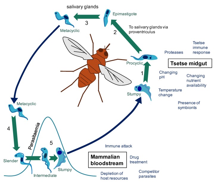
African trypanosomes cause devastating disease in sub-Saharan Africa in humans and livestock. The parasite lives extracellularly within the bloodstream of mammalian hosts and is transmitted by blood-feeding tsetse flies. In the blood, trypanosomes exhibit two developmental forms: the slender form and the stumpy form. The slender form proliferates in the bloodstream, establishes the parasite numbers and avoids host immunity through antigenic variation. The stumpy form, in contrast, is non-proliferative and is adapted for transmission. Here, we overview the features of slender and stumpy form parasites in terms of their cytological and molecular characteristics and discuss how these contribute to their distinct biological functions. Thereafter, we describe the technical developments that have enabled recent discoveries that uncover how the slender to stumpy transition is enacted in molecular terms. Finally, we highlight new understanding of how control of the balance between slender and stumpy form parasites interfaces with other components of the infection dynamic of trypanosomes in their mammalian hosts. This interplay between the host environment and the parasite's developmental biology may expose new vulnerabilities to therapeutic attack or reveal where drug control may be thwarted by the biological complexity of the parasite's lifestyle.

Keywords: Trypanosoma brucei; differentiation; life-cycle; quorum sensing; stumpy form; transmission.


Conflict of interest statement

The authors declare no conflict of interest.

Figures

Figure 1
Figure 1
The changing environments of the T. brucei life cycle. (1) Following a tsetse fly blood meal on an infected mammal (blue arrow), T. brucei stumpy cells reaching the tsetse midgut differentiate to procyclic cells. (2) Development of T. brucei proceeds through the tsetse midgut to the proventriculus. (3) Differentiation to epimastigote cells is followed by production of mammalian-infective metacyclic cells in the salivary gland. (4) On transfer to a new mammalian host via a tsetse fly bite (blue arrow), metacyclic cells differentiate to bloodstream forms. (5) In the bloodstream, proliferative slender cells elevate the parasitaemia until accumulation of a density-dependent signal triggers differentiation to the cell-cycle arrested stumpy form (via an intermediate stage). The stumpy form of T. brucei faces a particular challenge in that it must survive long enough in the bloodstream to be transmitted, but it must also survive long enough in the tsetse midgut to differentiate to the next life cycle stage and establish infection. These different environments pose different challenges that must be overcome to enable the continuation of the parasite life cycle. For example, in the mammalian host (red section), parasites must evade immune attack to ensure parasite survival and avoid excessive exploitation of host resources to ensure the host survives long enough to enable successful transmission to the tsetse fly vector. On uptake to the tsetse fly midgut (orange section), the parasites face a shift in nutrient availability and temperature among other challenges. For a review of factors effecting establishment of tsetse fly midgut infection see [1].
Figure 2
Figure 2
Characteristics of a stumpy cell. A number of assays can be used to test the proportion of cells in a T. brucei bloodstream form population that are slender or stumpy. (1) Firstly, stumpy cells are clearly morphologically distinct from slender cells. The representative cells shown have been labelled with the DNA stain DAPI (white) to highlight the position of the nucleus and kinetoplast in each case. (2) Secondly, stumpy cells express the PAD1 protein on their surface [27]. PAD1 positive cells can be identified using the PAD1 antibody by immunofluorescence microscopy or Western blotting. PAD-based reporter assays have also been established for enzymatic or cytological quantitation. (3) Stumpy cells also have a more elaborated mitochondrion than slender cells [23] and this can be detected by incubating the cells with mitotracker dye [24] before fixing and visualising the cells with a fluorescence microscope. (4) During the cell cycle, T. brucei cells first segregate their replicated mitochondrial genome (kinetoplast, K) and then their nucleus (N) prior to cytokinesis to generate two daughter cells [71]. Stumpy cells are arrested in G1/G0 of the cell cycle and have 1 kinetoplast and 1 nucleus (1K1N) [37]. Thus, differentiation to a stumpy population is accompanied by an accumulation of 1K1N cells, and this can be detected by staining with an appropriate DNA dye (e.g., DAPI) and KN scoring by immunofluorescence microscopy or flow cytometry (a completely stumpy population will have a single dominant peak corresponding to G1/G0). (5) Finally, stumpy cells, unlike slender cells, are able to differentiate synchronously to procyclic cells in culture [41], when differentiation is triggered by incubating cells at 27 °C in SDM-79 medium containing 6 mM cis-aconitate. Differentiation to procyclic cells is indicated by expression of EP procyclin [72] and can be detected using an EP procyclin antibody and flow cytometry. Maximal expression of EP procyclin is detectable 3 h after exposure of stumpy cells to cis-aconitate [51].

References

    1. Dyer N.A., Rose C., Ejeh N.O., Acosta-Serrano A. Flying tryps: Survival and maturation of trypanosomes in tsetse flies. Trends Parasitol. 2013;29:188–196. doi: 10.1016/j.pt.2013.02.003. - DOI - PubMed
    1. Vickerman K. Antigenic variation in trypanosomes. Nature. 1978;273:613–617. doi: 10.1038/273613a0. - DOI - PubMed
    1. Fenn K., Matthews K.R. The cell biology of Trypanosoma brucei differentiation. Curr. Opin. Microbiol. 2007;10:539–546. doi: 10.1016/j.mib.2007.09.014. - DOI - PMC - PubMed
    1. MacGregor P., Szoor B., Savill N.J., Matthews K.R. Trypanosomal immune evasion, chronicity and transmission: An elegant balancing act. Nat. Rev. Microbiol. 2012;10:431–438. doi: 10.1038/nrmicro2779. - DOI - PMC - PubMed
    1. Matthews K.R., McCulloch R., Morrison L.J. The within-host dynamics of African trypanosome infections. Philos. Trans. R. Soc. Lond. B Biol. Sci. 2015;370 doi: 10.1098/rstb.2014.0288. - DOI - PMC - PubMed

LinkOut - more resources