. 2017 Jul 6;377(1):52-61.
doi: 10.1056/NEJMoa1615887. Epub 2017 Jun 28.

CD55 Deficiency, Early-Onset Protein-Losing Enteropathy, and Thrombosis

Affiliations

CD55 Deficiency, Early-Onset Protein-Losing Enteropathy, and Thrombosis

Ahmet Ozen et al. N Engl J Med. .

Abstract

Background: Studies of monogenic gastrointestinal diseases have revealed molecular pathways critical to gut homeostasis and enabled the development of targeted therapies.

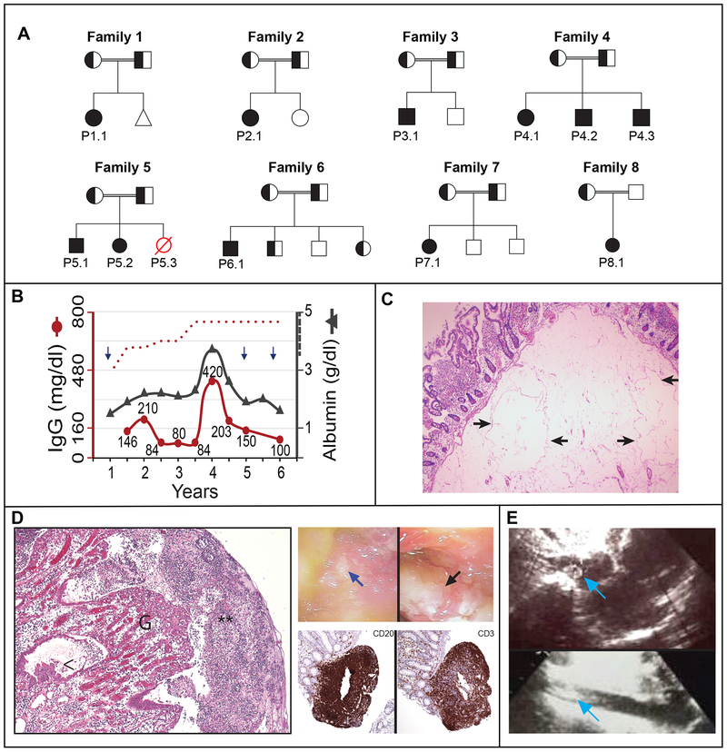
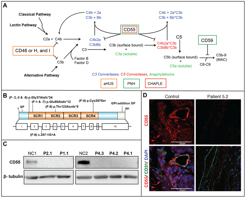
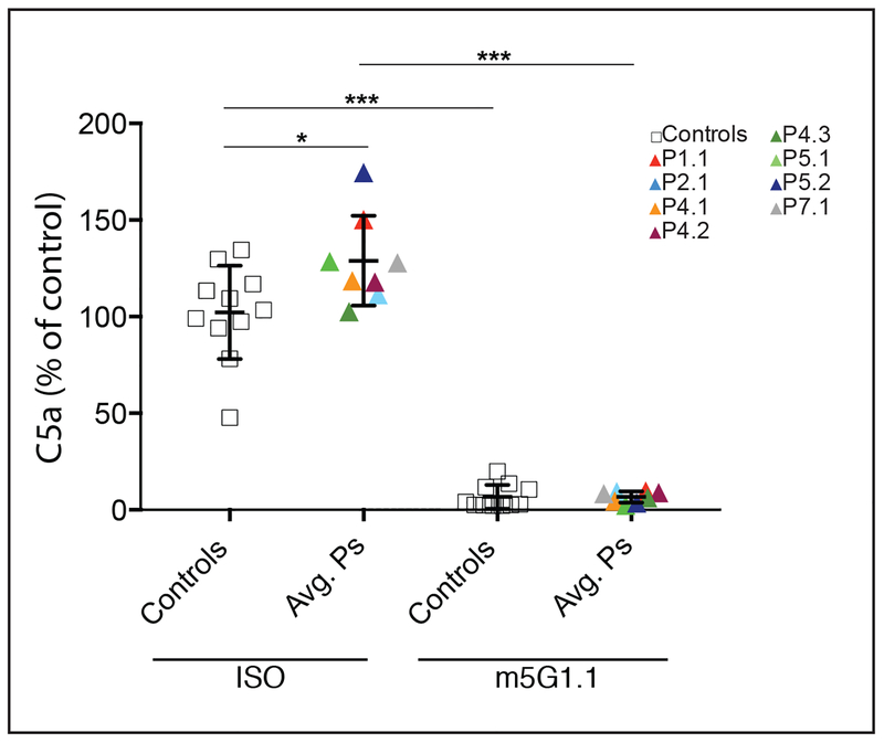
Methods: We studied 11 patients with abdominal pain and diarrhea caused by early-onset protein-losing enteropathy with primary intestinal lymphangiectasia, edema due to hypoproteinemia, malabsorption, and less frequently, bowel inflammation, recurrent infections, and angiopathic thromboembolic disease; the disorder followed an autosomal recessive pattern of inheritance. Whole-exome sequencing was performed to identify gene variants. We evaluated the function of CD55 in patients' cells, which we confirmed by means of exogenous induction of expression of CD55.

Results: We identified homozygous loss-of-function mutations in the gene encoding CD55 (decay-accelerating factor), which lead to loss of protein expression. Patients' T lymphocytes showed increased complement activation causing surface deposition of complement and the generation of soluble C5a. Costimulatory function and cytokine modulation by CD55 were defective. Genetic reconstitution of CD55 or treatment with a complement-inhibitory therapeutic antibody reversed abnormal complement activation.

Conclusions: CD55 deficiency with hyperactivation of complement, angiopathic thrombosis, and protein-losing enteropathy (the CHAPLE syndrome) is caused by abnormal complement activation due to biallelic loss-of-function mutations in CD55. (Funded by the National Institute of Allergy and Infectious Diseases and others.).


Figures

Figure 1.
Figure 1.. Clinical presentation of 8 families with familial early-onset protein-losing enteropathy
Panel A shows pedigrees of eight families with affected individuals homozygous for mutant allele indicated by solid symbols, heterozygous individuals indicated by half solid symbols, and affected individuals with an unknown genotype indicated by red, open symbols. The triangle in Family 1 represents a miscarriage. Panel B shows the serum levels of immunoglobulin G (IgG, left Y axis) in relation to serum albumin (right Y axis) concentrations as a function of age in years for Patient 1.1. Age-specific lower cutoff value for IgG is denoted by the red dotted curve, whereas the reference for albumin level is >3.5 g/dl (indicated by hatched line on right Y axis). Each arrow denotes an episode of pneumonia. Panel C shows H&E sections of resected duodenal tissue with markedly dilated lymphovascular spaces within submucosa in Patient 5.2 (arrows). Panel D (left) shows H&E sections from surgical resection material of small intestine which exhibits ulceration covered by fibrin with dense granulocytic infiltrate (**), granulation tissue (G) with edema in the lamina propria and reactive epithelial changes (<) in Patient 2.1 (left), as well as endoscopy photographs showing a mucosal ulcer (blue arrow) and exudate (black arrow) in the terminal ileum of Patient 1.1 (right, top) and histopathology of a biopsy of the ileum of Patient 2.1 and immunohistochemistry showing presence of B cells (CD20) and T cells (CD3) within lymphoid nodules (right, bottom). Panel E shows an echocardiographic image from Patient 6.1 with thrombus in the right atrium (arrow, top) and inferior vena cava (IVC) (arrow, bottom).
Figure 2.
Figure 2.. Variants in CD55 Lead to Loss of Protein Expression
Panel A shows the identified mutations relative to the CD55 protein structure depicting the four short consensus repeat (SCR1 – 4) domains (top) and exon structure (bottom) SP: signal peptide. Panel B shows a protein immunoblot for CD55 in activated CD4+ T cells. Panel C shows indirect immunofluorescence staining of duodenal biopsy taken from Patient 5.2 and healthy control staining for CD55 (red), the endothelial marker, CD31, (green) and DAPI (blue).
Figure 3.
Figure 3.. Loss of CD55 and Increased Complement Deposition
Panel A shows pooled analyses of C3d staining on T cells of five CD55-deficient patients after incubation with media (pH 7.4) containing pooled normal human serum (nHS) for the times indicated (right) HIS: heat inactivated serum. Panel B shows the pooled analysis of C3d staining with (+IgG1) or without (-IgG1) precoating with an anti-CD28 antibody to activate the classical pathway. Each color coded patient (P) data point represents the average (avg) of at least three repeats for a single patient. Panel C shows C3d staining in Patient 1.1 CD4+ T cells reconstituted with wild-type CD55 (left) or the control marker Thy1.1 (right) and then incubated with serum for 24 hours. Panel D shows C5b-9 staining of duodenal biopsies from a healthy donor and P5.2. In figure 3A the t-test was used to assess the significance between the two groups for the 5 hour incubation, while the nonparametric Mann–Whitney U-test was performed to assess the significance for the 1-hr, 2-hr and 24-hr conditions. In figure 3B the Mann–Whitney U-test was performed for comparisons between the control and patient groups and the Wilcoxon matched pairs signed rank test was applied to comparisons of the same group with or without treatment. (Isotype control (C), not significant (n.s.), *p<.05, **p<.01, ***p<.001).
Figure 4.
Figure 4.. Effect of Eculizumab on C5a production on Patient T Cells
Graph shows the levels C5a in supernatants of CD4+ T cell cultures, color coded by patient, after 2 hours of incubation with 10% normal human plasma (nHP) and 10 μg/mL of either isotype control (ISO) or C5 inhibitory (m5G1.1) antibodies. Each triangle represents the average of at least three repeat measurements for a single donor or patient. A two-tailed unpaired T test with Welch’s correction was performed for comparisons between the control and patient groups and the two tailed paired T test was applied to comparisons of the same group with or without treatment (not significant (n.s.), *p<.05, **p<.01, ***p<.001).

Comment in

Similar articles

Cited by

References

    1. McGovern DP, Kugathasan S, Cho JH. Genetics of Inflammatory Bowel Diseases. Gastroenterology 2015;149:1163–76 e2. - PMC - PubMed
    1. Umar SB, DiBaise JK. Protein-losing enteropathy: case illustrations and clinical review. Am J Gastroenterol 2010;105:43–9; quiz 50. - PubMed
    1. Elkadri A, Thoeni C, Deharvengt SJ, et al. Mutations in Plasmalemma Vesicle Associated Protein Result in Sieving Protein-Losing Enteropathy Characterized by Hypoproteinemia, Hypoalbuminemia, and Hypertriglyceridemia. Cell Mol Gastroenterol Hepatol 2015;1:381–94 e7. - PMC - PubMed
    1. Waldmann TA, Steinfeld JL, Dutcher TF, Davidson JD, Gordon RS Jr., The role of the gastrointestinal system in “idiopathic hypoproteinemia”. Gastroenterology 1961;41:197–207. - PubMed
    1. Alders M, Hogan BM, Gjini E, et al. Mutations in CCBE1 cause generalized lymph vessel dysplasia in humans. Nature genetics 2009;41:1272–4. - PubMed

MeSH terms