

Genetic basis of calcifying cystic odontogenic tumors
- PMID: 28658279
- PMCID: PMC5489209
- DOI: 10.1371/journal.pone.0180224
Genetic basis of calcifying cystic odontogenic tumors
Abstract
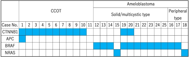
Calcifying cystic odontogenic tumors (CCOTs) are benign cystic tumors that form abnormally keratinized ghost cells. Mutations in CTNNB1, which encodes beta-catenin, have been implicated in the development of these tumors, but a causal relationship has not been definitively established. Thus, mutational hot spots in 50 cancer genes were examined by targeted next-generation sequencing in 11 samples of CCOT. Mutations in CTNNB1, but not in other genes, were observed in 10 of 11 cases. These mutations constitutively activate beta-catenin signaling by abolishing the phosphorylation sites Asp32, Ser33, or Ser37, and are similar to those reported in pilomatrixoma and adamantinomatous craniopharyngioma. In contrast, BRAF or NRAS mutations were observed in 12 and two control samples of ameloblastoma, respectively. In HEK293 cells, overexpression of mutated CTNNB1 also upregulated hair keratin, a marker of ghost cells. Furthermore, ghost cells were present in two cases of ameloblastoma with BRAF and CTNNB1 mutations, indicating that ghost cells form due to mutations in CTNNB1. The data suggest that mutations in CTNNB1 are the major driver mutations of CCOT, and that CCOT is the genetic analog of pilomatrixoma and adamantinomatous craniopharyngioma in odontogenic tissue.
Conflict of interest statement
Figures


References
-
- Barnes L, Eveson JW, Reichart P, Sidransky D. Pathology & Genetics Head and Neck Tumors: IARC Press; 2005
-
- Robinson RA, Vincent SD. Tumors and Cysts of the Jaws: American registry of Pathology; 2012.
-
- Rajesh E, Jimson S, Masthan KM, Balachander N. Ghost cell lesions. J Pharm Bioallied Sci. 2015;7(Suppl 1): S142–144. doi: 10.4103/0975-7406.155864 - DOI - PMC - PubMed
-
- Ledesma-Montes C, Gorlin RJ, Shear M, Prae Torius F, Mosqueda-Taylor A, Altini M, et al. International collaborative study on ghost cell odontogenic tumours: calcifying cystic odontogenic tumour, dentinogenic ghost cell tumour and ghost cell odontogenic carcinoma. J Oral Pathol Med. 2008;37(5):302–308. doi: 10.1111/j.1600-0714.2007.00623.x - DOI - PubMed
-
- Praetorius F, Hjorting-Hansen E, Gorlin RJ, Vickers RA. Calcifying odontogenic cyst. Range, variations and neoplastic potential. Acta Odontol Scand. 1981;39(4):227–240. - PubMed
MeSH terms
Substances
LinkOut - more resources
Full Text Sources
Other Literature Sources
Molecular Biology Databases
Research Materials
Miscellaneous
