

Abnormal dendritic maturation of developing cortical neurons exposed to corticotropin releasing hormone (CRH): Insights into effects of prenatal adversity?
- PMID: 28658297
- PMCID: PMC5489219
- DOI: 10.1371/journal.pone.0180311
Abnormal dendritic maturation of developing cortical neurons exposed to corticotropin releasing hormone (CRH): Insights into effects of prenatal adversity?
Abstract
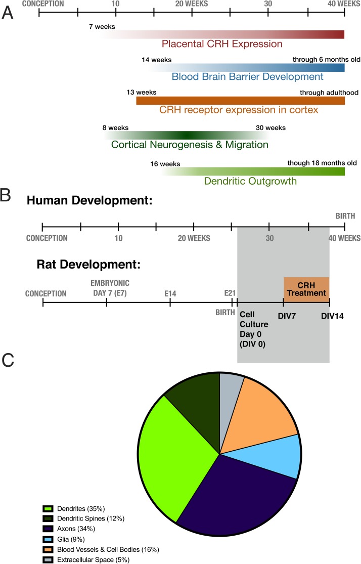
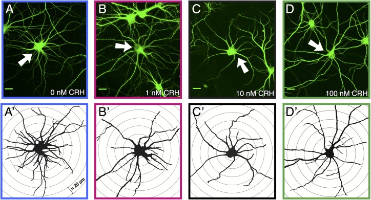
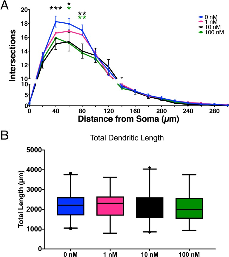
Corticotropin releasing hormone (CRH) produced by the hypothalamus initiates the hypothalamic-pituitary-adrenal (HPA) axis, which regulates the body's stress response. CRH levels typically are undetectable in human plasma, but during pregnancy the primate placenta synthesizes and releases large amounts of CRH into both maternal and fetal circulations. Notably, placental CRH synthesis increases in response to maternal stress signals. There is evidence that human fetal exposure to high concentrations of placental CRH is associated with behavioral consequences during infancy and into childhood, however the direct effects on of the peptide on the human brain are unknown. In this study, we used a rodent model to test the plausibility that CRH has direct effects on the developing cortex. Because chronic exposure to CRH reduces dendritic branching in hippocampal neurons, we tested the hypothesis that exposure to CRH would provoke impoverishment of dendritic trees in cortical neurons. This might be reflected in humans as cortical thinning. We grew developing cortical neurons in primary cultures in the presence of graded concentrations of CRH. We then employed Sholl analyses to measure dendritic branching and total dendritic length of treated cells. A seven-day exposure to increasing levels of CRH led to a significant, dose-dependent impoverishment of the branching of pyramidal-like cortical neurons. These results are consistent with the hypothesis that, rather than merely being a marker of prenatal stress, CRH directly decreases dendritic branching. Because dendrites comprise a large portion of cortical volume these findings might underlie reduced cortical thickness and could contribute to the behavioral consequences observed in children exposed to high levels of CRH in utero.
Conflict of interest statement
Figures
References
-
- Vale W, Spiess J, Rivier C, Rivier J. Characterization of a 41-residue ovine hypothalamic peptide that stimulates secretion of corticotropin and beta-endorphin. Science (80-). 1981. September 18;213(4514):1394–7. - PubMed
-
- Bale TL, Vale WW. CRF AND CRF R ECEPTORS: Role in Stress Responsivity and Other Behaviors. Annu Rev Pharmacol Toxicol. 2004. February 10;44(1):525–57. - PubMed
-
- Herman JP, McKlveen JM, Ghosal S, Kopp B, Wulsin A, Makinson R, et al. Regulation of the Hypothalamic-Pituitary-Adrenocortical Stress Response In: Comprehensive Physiology. Hoboken, NJ, USA: John Wiley & Sons, Inc.; 2016. p. 603–21. doi: 10.1002/cphy.c150015 - DOI - PMC - PubMed
-
- Regev L, Baram TZ. Corticotropin releasing factor in neuroplasticity. Front Neuroendocrinol. 2014. April;35(2):171–9. doi: 10.1016/j.yfrne.2013.10.001 - DOI - PMC - PubMed
MeSH terms
Substances
Grants and funding
LinkOut - more resources
Full Text Sources
Other Literature Sources
