. 2017 Jun 28;12(6):e0180097.
doi: 10.1371/journal.pone.0180097. eCollection 2017.

Critical role of SIK3 in mediating high salt and IL-17 synergy leading to breast cancer cell proliferation

Affiliations

Critical role of SIK3 in mediating high salt and IL-17 synergy leading to breast cancer cell proliferation

Suneetha Amara et al. PLoS One. .

Abstract

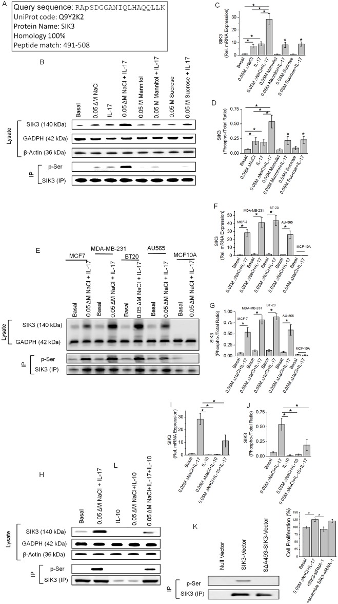
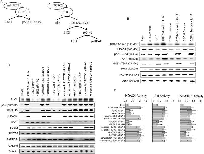
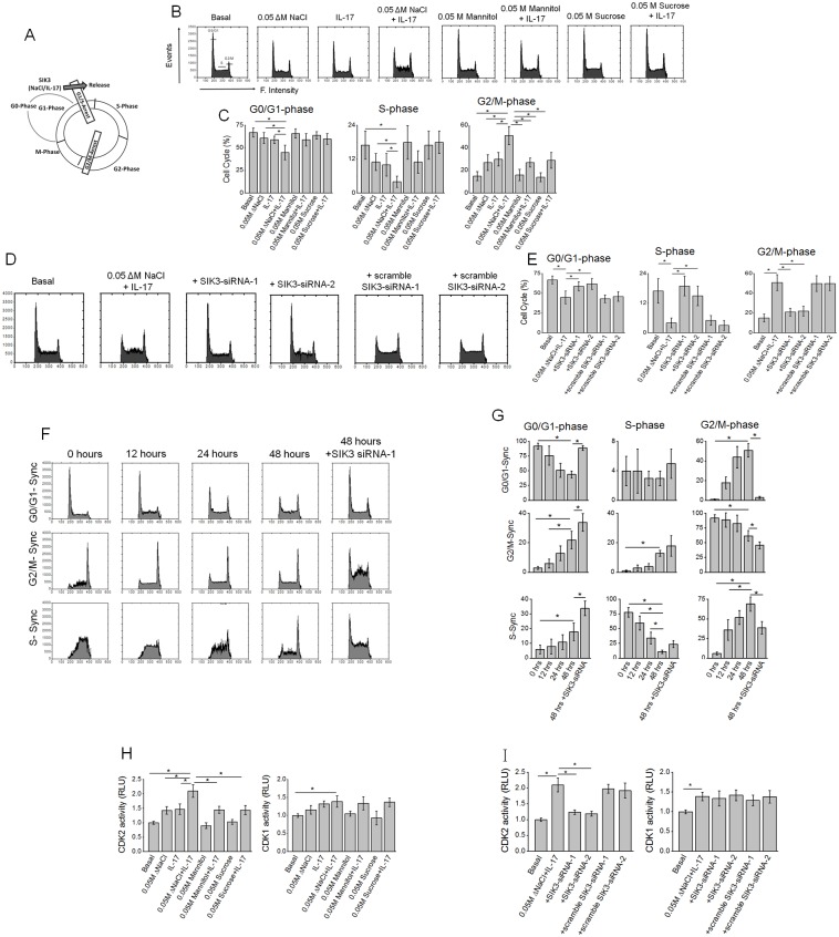
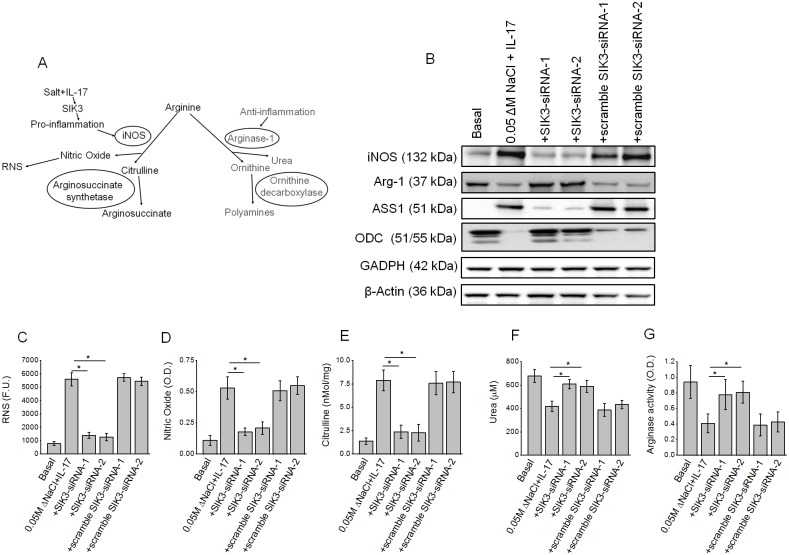
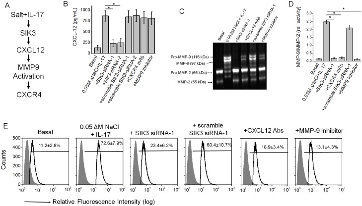
Chronic inflammation is a well-known precursor for cancer development and proliferation. We have recently demonstrated that high salt (NaCl) synergizes with sub-effective interleukin (IL)-17 to induce breast cancer cell proliferation. However, the exact molecular mechanisms mediating this effect are unclear. In our current study, we adopted a phosphoproteomic-based approach to identify salt modulated kinase-proteome specific molecular targets. The phosphoprotemics based binary comparison between heavy labelled MCF-7 cells treated with high salt (Δ0.05 M NaCl) and light labelled MCF-7 cells cultured under basal conditions demonstrated an enhanced phosphorylation of Serine-493 of SIK3 protein. The mRNA transcript and protein expression analysis of SIK3 in MCF-7 cells demonstrated a synergistic enhancement following co-treatment with high salt and sub-effective IL-17 (0.1 ng/mL), as compared to either treatments alone. A similar increase in SIK3 expression was observed in other breast cancer cell lines, MDA-MB-231, BT20, and AU565, while non-malignant breast epithelial cell line, MCF10A, did not induce SIK3 expression under similar conditions. Biochemical studies revealed mTORC2 acted as upstream mediator of SIK3 phosphorylation. Importantly, cell cycle analysis by flow cytometry demonstrated SIK3 induced G0/G1-phase release mediated cell proliferation, while SIK3 silencing abolished this effect. Also, SIK3 induced pro-inflammatory arginine metabolism, as evidenced by upregulation of the enzymes iNOS and ASS-1, along with downregulation of anti-inflammatory enzymes, arginase-1 and ornithine decarboxylase. Furthermore, gelatin zymography analysis has demonstrated that SIK3 induced expression of tumor metastatic CXCR4 through MMP-9 activation. Taken together, our data suggests a critical role of SIK3 in mediating three important hallmarks of cancer namely, cell proliferation, inflammation and metastasis. These studies provide a mechanistic basis for the future utilization of SIK3 as a key drug discovery target to improve breast cancer therapy.


Conflict of interest statement

Competing Interests: There is no conflict of interest for any of the authors regarding the submission and publication of this manuscript.

Figures

Fig 1
Fig 1. Identification of upregulation of SIK3 specifically following a high salt synergized IL-17 stimulation of breast cancer cells.
(A) Identification of the 1.9 fold higher enriched phospho-peptide sequence following high salt induced stimulation of MCF-7 cells. The identified sequence demonstrated 100% homology to the SIK3 protein at the serine-493 residue. (B) Western blot analysis of SIK3 expression in total cell lysate. Immunoprecipitation of SIK3 is probed with phospho-serine antibody. As can be noted, SIK3 expression was upregulated following treatment with high salt (0.05 M NaCl) and IL-17 (0.1 ng/mL) individually, further, SIK3 expression was synergistically elevated following co-treatment with high salt and IL-17. Equimolar mannitol (0.05 M) and sucrose (0.05 M) were used as negative controls. (C) mRNA transcript expression of SIK3 by ΔΔcT method of quantitative real time polymerase chain reaction normalized for GADPH demonstrated a significant upregulation following high salt (7.3±1.4 fold) and IL-17 (9.1±1.7 fold), and co-treatment with both high salt and IL-17 induced a (28.6±4.4 fold) synergistically enhanced expression. (D) Densitometry quantitation of phospho-SIK3 in (B) demonstrated a synergistically enhanced phosphorylation of SIK3 by high salt and IL-17. (E-G) Expression of SIK3 following co-treatment with high salt and IL-17 in five breast tissue related cell lines (E) were used in our studies, of these, four breast cancer cells (MCF7, MDA-MB-231, BT20, AU565) and one non-malignant breast epithelial cell line (MCF10A); mRNA transcript analysis in 5 cell lines (F); densitometry quantitation of phosphorylated SIK3 in five cell lines (G). (H-J) Anti-inflammatory cytokine interleukin-10 (IL-10) inhibited the expression of SIK3 protein (H), mRNA transcript (I) and phosphorylation of SIK3(J). (K) To demonstrate the phosphorylation is specifically on Serine-493 of SIk3 we clone the SIK3 into MCF10A cell line and latter stimulated by co-treatment with high salt and IL-17. Of note, wild type MCF10A did not demonstrate SIK3 expression following co-treatment with high salt and IL-17 (E-G). Mutation of serine to alanine (SΔA493-SIK3) did not demonstration any binding with phospho-serine in SIK3 immunoprecipitate. (L) Cell proliferation of MCF-7 cells was inhibited following SIK3 siRNA treatment following co-treatment with high salt and IL-17. Of note, we have previously demonstrated that (Ref [6, 13]) co-treatment with high salt and IL-17 induced a 25% higher proliferation of MCF-7 cancer cells. The other three cancer cells MDA-MB-231 (29% higher), BT-20 (31% higher) and AU565 (24% higher) cell proliferation following co-treatment with high salt and IL-17, while non-malignant breast epithelial MCF10A cells did not demonstrate any enhanced proliferation following similar treatment conditions. All data represented as mean values ± SEM from four independent experiments. Student-t-test performed for statistical analysis (significance p<0.05).
Fig 2
Fig 2. mTORC2 is an upstream upregulator of SIK3 expression and phosphorylation.
(A) Schematic of the potential interplay between mTOR2 and SIK3. (B) Phosphorylation of HDAC4, AKT, and S6K1 the downstream targets for SIK3, mTOR1 and mTOR2, respectively, were analyzed following stimulation with high salt and IL-17, individually and combined by Western blot. (C) Phosphorylation of HDAC4, AKT, and S6K1 the downstream targets for SIK3, mTOR1 and mTOR2, respectively, were analyzed by Western blot following co-treatment with high salt and IL-17, and Knock down of SIK3, RAPTOR and RICTOR. Scramble siRNA of the original construct were used as controls. (D) Activity of HDAC4, Akt and S6K1 were analyzed by ELISA under various conditions mentioned in (C). Of note, HDAC4 activity was decreased following SIK3 and RICTOR knock down, while, mTOR2 downstream target AKT and mTOR1 downstream target S6K1 activity was decreased following RICTOR and RAPTOR, respectively. All data represented as mean values ± SEM from four independent experiments. Student-t-test performed for statistical analysis (significance p<0.05).
Fig 3
Fig 3. High salt synergized with IL-17 to induce SIK3 mediated G0/G1-release in cell cycle.
(A) Schematic of cell cycle and potential impact of co-treatment with high salt and IL-17. (B) Flow cytometry analysis of cell cycle following treatment with high salt and IL-17, individually and combined. (C) Relative percentage of cells (%) in G0/G1-, S-, and G2/M-phase following various treatment conditions mentioned above. (D-E) Impact of SIK3 knockdown by siRNA on the three cell cycle phases. (F-G) Temporal Synchronization cell cycle studies following initial chemical synchronization to either G0/G1-, S-, or G2/M-phase and followed by co-treatment with high salt and IL-17 with cell cycle analysis done at 0, 12, 24 and 48 hour time points. The last panel is with cells initially synchronized to various cell cycle phases followed by co-treatment with high salt and IL-17 for 12 hours, followed by siRNA transfection (SIK3 or scramble) at 12 hours and followed by co-treatment with high salt and IL-17 for another 36 hours and data analyzed at 48 hour-time point. (H) Analysis of G0/G1-phase release mediating CDK2 and G2/M-phase release mediating CDK1 activity at 48 hour time point following treatment with high salt and IL-17 individually and combined. As can be noted, co-treatment with high salt and IL-17 induced CDK2 activity and release from G0/G1-phase arrest of cell cycle. (I) Analysis of G0/G1-phase release mediating CDK2 and G2/M-phase release mediating CDK1 activity following siRNA knockdown of SIK3 (as mentioned under F-G)and co-treatment with high salt and IL-17. All data represented as mean values ± SEM from four independent experiments. Student-t-test performed for statistical analysis (significance p<0.05).
Fig 4
Fig 4. High salt synergized with IL-17 to induce SIK3 mediated pro-inflammatory nitric oxide release components of arginine metabolism while suppressing anti-inflammatory components of arginine metabolism.
(A) Schematic of arginine metabolism under potential pro-inflammatory and anti-inflammatory stimulus conditions. (B) Western blot analysis of the pro-inflammation mediating enzymes inducible nitric oxide synthetase (iNOS) and arginosuccinate synthetase (ASS-1); and anti-inflammation mediating enzymes arginase-1 (Arg-1) and ornithine decarboxylase (ODC) following co-treatment with high salt and IL-17 along with SIK3 knockdown. (C-G) production of reactive nitrogen species (C), nitric oxide (D), citrulline (E), urea (F) and arginase activity (G) following co-treatment with high salt and IL-17, along with SIK3 knock down. All data represented as mean values ± SEM from four independent experiments. Student-t-test performed for statistical analysis (significance p<0.05).
Fig 5
Fig 5. High salt synergized with IL-17 to induced SIK3 mediated expression of tumor metastatic CXCR4 through MMP-9 activation.
(A) Schematic of the metastasis mediating CXCL12/CXR4. (B) ELISA-based analysis in the cell supernatant for the CXCL12 expression following co-treatment with high salt and IL-17 along with SIK3 knockdown or MMP-9 inhibitor (10 μM, 2-(N-Benzyl-4-methoxyphenylsulfonamido)-5-((diethylamino)methyl)-N-hydroxy-3-methylbenzamide, ab142180, AbCam, Cambridge, MA) or CXCR4 blocked with specific monoclonal antibody (Santa Cruz, Dallas, TX). (C) Gelatin Zymography to analysis the MMP-9/-2 activity under the above mentioned treatment conditions. (D) Ratio of inducible MMP-9 to constitutive MMP-2 activity under above mentioned treatment conditions. (E) Flow cytometry-based analysis in the membrane localization of the CXCR-4 following co-treatment with high salt and IL-17 along with SIK3 knockdown or MMP-9 inhibitor or CXCL12 blocked with specific monoclonal antibody (Santa Cruz, Dallas, TX). All data represented as mean values ± SEM from four independent experiments. Student-t-test performed for statistical analysis (significance p<0.05).

References

    1. Crusz SM, Balkwill FR. Inflammation and cancer: advances and new agents. Nat Rev Clin Oncol 2015; 12(10): 584–596. doi: 10.1038/nrclinonc.2015.105 - DOI - PubMed
    1. Hanahan D, Weinberg RA. Hallmarks of cancer: the next generation. Cell 2011; 144(5): 646–674. doi: 10.1016/j.cell.2011.02.013 - DOI - PubMed
    1. Landskron G, De la Fuente M, Thuwajit P, Thuwajit C, Hermoso MA. Chronic inflammation and cytokines in the tumor microenvironment. J Immunol Res 2014; 2014: 149185 doi: 10.1155/2014/149185 - DOI - PMC - PubMed
    1. Bailey SR, Nelson MH, Himes RA, Li Z, Mehrotra S, Paulos CM. Th17 cells in cancer: the ultimate identity crisis. Front Immunol 2014; 5: 276 doi: 10.3389/fimmu.2014.00276 - DOI - PMC - PubMed
    1. Wu C, Yosef N, Thalhamer T, Zhu C, Xiao S, Kishi Y et al. Induction of pathogenic TH17 cells by inducible salt-sensing kinase SGK1. Nature 2013; 496(7446): 513–517. doi: 10.1038/nature11984 - DOI - PMC - PubMed