. 2017 Jun 28;5(1):53.
doi: 10.1186/s40478-017-0456-2.

The novel compound PBT434 prevents iron mediated neurodegeneration and alpha-synuclein toxicity in multiple models of Parkinson's disease

Affiliations

The novel compound PBT434 prevents iron mediated neurodegeneration and alpha-synuclein toxicity in multiple models of Parkinson's disease

David I Finkelstein et al. Acta Neuropathol Commun. .

Erratum in

Abstract

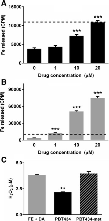
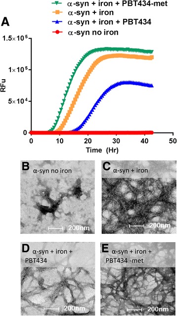
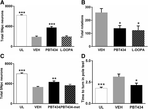
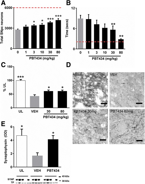
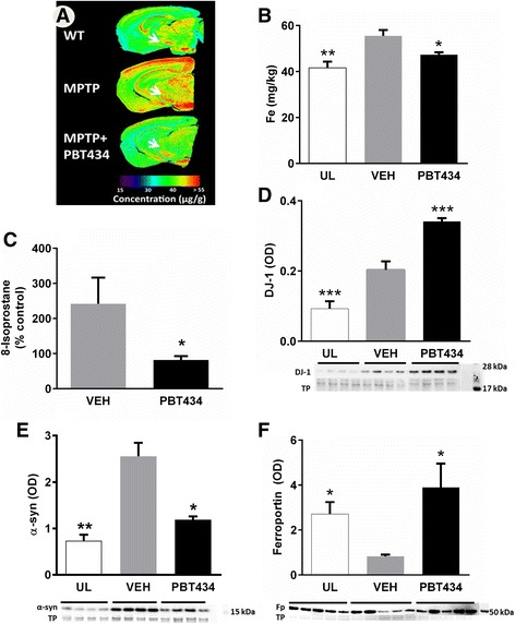
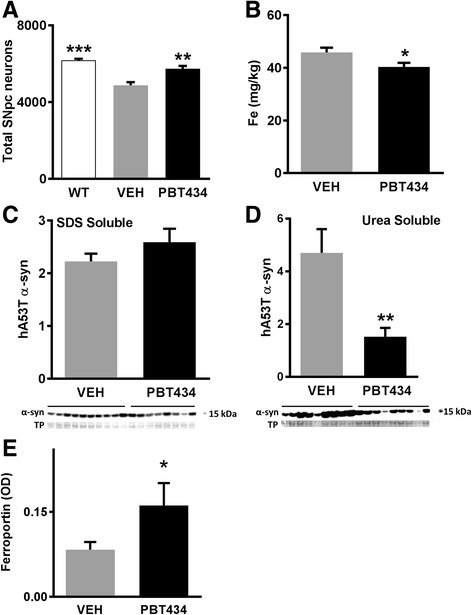
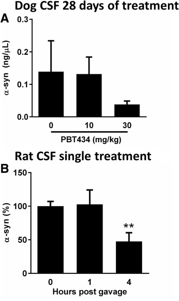
Elevated iron in the SNpc may play a key role in Parkinson's disease (PD) neurodegeneration since drug candidates with high iron affinity rescue PD animal models, and one candidate, deferirpone, has shown efficacy recently in a phase two clinical trial. However, strong iron chelators may perturb essential iron metabolism, and it is not yet known whether the damage associated with iron is mediated by a tightly bound (eg ferritin) or lower-affinity, labile, iron pool. Here we report the preclinical characterization of PBT434, a novel quinazolinone compound bearing a moderate affinity metal-binding motif, which is in development for Parkinsonian conditions. In vitro, PBT434 was far less potent than deferiprone or deferoxamine at lowering cellular iron levels, yet was found to inhibit iron-mediated redox activity and iron-mediated aggregation of α-synuclein, a protein that aggregates in the neuropathology. In vivo, PBT434 did not deplete tissue iron stores in normal rodents, yet prevented loss of substantia nigra pars compacta neurons (SNpc), lowered nigral α-synuclein accumulation, and rescued motor performance in mice exposed to the Parkinsonian toxins 6-OHDA and MPTP, and in a transgenic animal model (hA53T α-synuclein) of PD. These improvements were associated with reduced markers of oxidative damage, and increased levels of ferroportin (an iron exporter) and DJ-1. We conclude that compounds designed to target a pool of pathological iron that is not held in high-affinity complexes in the tissue can maintain the survival of SNpc neurons and could be disease-modifying in PD.

Keywords: Chelation; Drug development; Neuroprotection; Oxidative stress; Synucleinopathy.


Figures

Fig. 1
Fig. 1
PBT434 enhances the release of iron and prevents the generation of hydrogen peroxide. Cultured M17 neuroblastoma cells were loaded with the iron isotope 59Fe. The cells were washed and then exposed to a chelator to assess if iron could be removed from the cell. Cells loaded with the iron isotope 59Fe were exposed to a PBT434 and the amount of radioactive 59Fe released into the media was measured (CPM = counts per minute) or b deferiprone at 0, 1, 10 or 20 μM for 3 h. Deferiprone showed a dose related increase in the levels of 59Fe secreted into growth medium. With PBT434 the effect was observed only at the highest dose of 20 μM (*P < 0.05, ** P < 0.01, *** P < 0.0001, One-way ANOVA, Tukey Post Hoc). At the highest concentration, the effect of deferiprone was 5-fold greater than for PBT434. The dashed line represents equivalent values on the two graphs. c PBT434 causes an inhibition of metal mediated redox activity. Fe-citrate (0.4 μM) in the presence of dopamine (DA, 50 mM) generate hydrogen peroxide (H2O2) assessed using a cell-free fluorescence-based assay. PBT434 at 10 μM but not PBT434-met significantly reduced H2O2 generated by Fe/DA (PBT434-met = analog of PBT434 in which the metal binding site is blocked; One-way ANOVA, Tukey Post Hoc). Dopamine without Fe-Citrate did not produce H2O2
Fig. 2
Fig. 2
Inhibition of iron mediated α-synuclein (α-syn) aggregation. Recombinant α-synuclein (186.6 μM) was incubated alone or in the presence of equimolar concentrations of iron nitrate (iron = Fe (NO3)3), PBT434 or PBT434-met. Thioflavin T (ThT) fluorescence was measured (RFU = relative fluorescent units) every 30 min for 42 h. The lag-time of aggregation was slowed by PBT434 (α-synuclein = 37 h, α-syn + iron = 10.2 h, α-syn + iron + PBT434 = 16.60 h and α-syn + iron + PBT434-met =7.9 h). a One Way ANOVA with games-howell post hoc analysis (unequal variances on the lag-phases, individual reaction wells (n = 5–6) showed that, α-syn + iron had significantly faster aggregation than α-syn + iron + PBT434 (p = 0.02) or α-syn alone (p = 0.04) but not α-syn + iron + PBT434-met (p = 0.49). Lower panels: Electron micrographs of samples from each reaction mixture were taken after 42 h (b; α- synuclein, no iron); c Increased fibril formation in the presence of iron; d Diminished fibril formation in the presence of PBT434; E) PBT434-met did not reduce the fibril formation
Fig. 3
Fig. 3
PBT434 prevents toxin induced cell loss and improves motor performance. a 6-OHDA injection resulted in the loss of 65% of the SNpc neurons compared with unlesioned control animals. PBT434 administration commencing 3 days following intoxication and significantly preserved neuron numbers compared with vehicle (p < 0.001, One-way ANOVA, Tukey Post Hoc). The number of neurons in an unlesioned mouse is 6124 ± 23. L-DOPA did not protect neurons against 6-OHDA toxicity. b Mice treated with PBT434 (30 mg/kg/day, N = 11 (P < 0.05) or L-DOPA (15 mg/kg, P < 0.001, One-way ANOVA, Tukey Post Hoc) showed significantly fewer rotations than vehicle treated mice. c PBT434 (30 mg/kg/day for twenty days) administered 24 h following intoxication with MPTP resulted in significantly reduced SNpc neuronal loss (*** P < 0.001, One-way ANOVA, Tukey Post Hoc). PBT434-met 30 mg/kg/day (PBT434 without the metal binding site) does not protect against MPTP. d PBT434 treatment resulted in improvement in motor performance in the Pole test (* P < 0.05, One-way ANOVA, Tukey Post Hoc). UL = unlesioned, VEH = standard suspension vehicle without compound, PBT434-met = analogue of PBT434 without the metal binding
Fig. 4
Fig. 4
Dose response effects of PBT434 on neuron number and TH - positive varicosities. The effects on SNpc neuron number and motor function in response to escalating dose of PBT434 were assessed. 12–14 week old male C57BL/6 mice were lesioned using MPTP (60 mg/kg) resulting in an average SNpc lesion size of 55%. Treatment with PBT434 at 1,3,10, 30 or 80 mg/kg/day commenced 24 h after induction of lesion. a Mean number of SNpc neurons compared with the vehicle treated group (VEH)(±SEM). The proportion of SNpc cells rescued increased with increasing dose of PBT434. 3 mg (P < 0.05), 10 mg (P < 0.01), 30 mg (P < 0.001) and 80 mg/kg/day (P < 0.001) doses prevented a significant proportion of cell loss by day 21 (73 animals were studied from two separate MPTP experiments; One-way ANOVA paired with Games-Howell post hoc test). The red dotted line indicates the normal value. b Pole test was undertaken at day 20 post intoxication to test motor performance. As the dose of PBT434 increased there was a trend to improved turning behavior compared with the vehicle group while doses 30 mg/kg/day (P < 0.05) and 80 mg/kg/day (P < 0.01) showed a significant 2.5–3 fold improvement in the time to turn. (One-way ANOVA paired with Games-Howell post hoc test). The dashed line represents the average time taken by unlesioned animals to perform the task. c The abundance of tyrosine hydroxylase-positive varicosities in the caudate putamen was assessed by stereology at day 21 following MPTP (N = 6–7 animals per treatment). Following MPTP intoxication, TH -positive varicosities were reduced compared with unlesioned mice (* p < 0.05. One-way ANOVA, Tukey’s Post hoc comparison). Treatment with 30 or 80 mg/kg of PBT434 significantly increased varicosity abundance compared with untreated mice. d Light micrographs of the dorso-lateral tier of the caudate putamen showing individual tyrosine hydroxylase -positive varicosities (arrows), for unlesioned animals (UL), MPTP lesioned vehicle treated (VEH) or following PBT434 treatment (scale bar = 25 μm). e Western blots of the dorsal tier of the caudate putamen showed that treatment with PBT434 prevented the decline in levels of the presynaptic marker synaptophysin (SYNP), * p < 0.05, One-way ANOVA, Tukey Post hoc comparison). TP = total protein, OD = optical density
Fig. 5
Fig. 5
PBT434 improves iron level following MPTP lesion. 12–14 week old male C57BL/6 mice were lesioned using MPTP which resulted in a lesion size 65–70% cell loss by day 21. MPTP lesioned mice were treated with vehicle (VEH) or PBT434 (30 mg/kg/day) from day 1 to day 21. a Brain samples collected at day 21 were sectioned and scanned using laser ablation-inductively coupled plasma-mass spectrometry. Representative images show Fe distribution in normal, unlesioned, wildtype (C57BL6) mouse brain, MPTP lesioned brain and MPTP + PBT434 treated brain. The heat map quantifies the level of iron in the SN, which is indicated by the arrow. b The concentrations of Fe (mg/kg) in the substantia nigra (SN, includes both compacta and reticulata) of the groups of mice were plotted onto a bar graph. MPTP injury causes a significant elevation in Fe in the SN at day 21 (**p < 0.01, one-way ANOVA, Tukey post hoc) compared with unlesioned mice (UL) which was attenuated by PBT434 (* p < 0.05, one-way ANOVA, Tukey post hoc). c PBT434 significantly prevented the MPTP induced elevation of 8-isoprostane within SN as measured by ELISA (*P < 0.05, One-way ANOVA, Tukey Post Hoc). d Western blot was used to measure levels of levels of DJ-1 in the SN. Levels of DJ-1 were significantly elevated with MPTP treatment in the absence of drug (VEH) and significantly further elevated with PBT434 (TP = Total Protein; OD = optical density; ***P < 0.001, One-way ANOVA, Tukey post hoc). The protein ran at the predicted molecular weight (24 kDa) as can be seen by comparing the position of the proteins with the molecular weight ladder on the right of the image. e α-synuclein levels in mice administered MPTP or MPTP + PBT434 (30 mg/kg/day, were compared with unlesioned controls; UL). SN tissue samples were homogenized to form a lysate, which was assayed by Western blot and quantitated by optical density (OD) normalized to total protein (TP, Ponceau). The protein ran at the predicted molecular weight 14 kDa compared to with the molecular weight ladder on the right of the image. In MPTP lesioned mice α-synuclein was significantly elevated by day 21 (**P < 001, one-way ANOVA, Tukey post hoc). α-synuclein protein levels were significantly lower with PBT434 treatment (*P < 0.05, one-way ANOVA, Tukey post hoc) compared with Vehicle treated animals. f Western blot of MPTP lesioned mice showed a significant reduction in levels of ferroportin protein which were decreased 21 days after the lesion (*P < 0.05). The protein ran at slightly less the predicted molecular weight 63 kDa compared to with the molecular weight ladder on the right of the image. Ferroportin protein levels were significantly higher with PBT434 treatment (*P < 0.05) compared with the vehicle treated animals but not different to unlesioned mice (one-way ANOVA, Tukey post hoc)
Fig. 6
Fig. 6
PBT434 modulates α-synuclein transgenic animals. hA53T α-synuclein Tg mice consumed an average of 37 mg/kg/day in animal chow of PBT434 from 4 months of age for 4 months. a PBT434 preserved SNpc neurons (** P < 0.01, one-way ANOVA, Tukey post hoc); b PBT434 decreased SN iron measured by mass spectrometry (*P < 0.05, T-Test). SN tissue samples were homogenized to form a lysate, which was assayed by western blot and quantitated by optical density (OD) normalized to total protein (TP, Ponceau). c PBT434 did not reduced levels of the SDS soluble fraction of α- synuclein in the SN (Western blot, urea soluble fraction); d PBT434 reduced levels of the urea soluble fraction of α- synuclein in the SN (** p < 0.01; T-Test); E) PBT434 treatment increased SN ferroportin levels (*P < 0.05, T-Test)
Fig. 7
Fig. 7
α-synuclein levels in CSF of dogs and rats following PBT434 treatment. α-synuclein was collected and quantified from CSF of dogs and rats following PBT434 treatment. a α-synuclein levels of CSF collected from dogs following 28 days exposure to PBT434 at various doses. α-synuclein levels detected by enhanced Western blot decline in the 10 mg/kg, did not reach significance (one-way ANOVA, Tukey post hoc). b Cannula were implanted into the lateral ventricles of wild type rats and CSF was sampled before and after gavage with 30 mg/kg/day PBT434. Western blot for the presence of showed a significant decrease in α-synuclein at 4 h but not at 1 h (* P < 0.05, one-way ANOVA, Tukey post hoc)

References

    1. Adlard PA, Cherny RA, Finkelstein DI, Gautier E, Robb E, Cortes M, Volitakis I, Liu X, Smith JP, Perez K, et al. Rapid restoration of cognition in Alzheimer’s transgenic mice with 8-hydroxy quinoline analogs is associated with decreased interstitial Abeta. Neuron. 2008;59:43–55. doi: 10.1016/j.neuron.2008.06.018. - DOI - PubMed
    1. Adlard PA, Bica L, White AR, Nurjono M, Filiz G, Crouch PJ, Donnelly PS, Cappai R, Finkelstein DI, Bush AI. Metal ionophore treatment restores dendritic spine density and synaptic protein levels in a mouse model of Alzheimer’s disease. PLoS One. 2011;6:e17669. doi: 10.1371/journal.pone.0017669. - DOI - PMC - PubMed
    1. Aisen P, Leibman A, Zweier J. Stoichiometric and site characteristics of the binding of iron to human transferrin. J Biol Chem. 1978;253:1930–1937. - PubMed
    1. Ayton S, Lei P, Duce JA, Wong BX, Sedjahtera A, Adlard PA, Bush AI, Finkelstein DI. Ceruloplasmin dysfunction and therapeutic potential for Parkinson disease. Ann Neurol. 2013;73:554–559. doi: 10.1002/ana.23817. - DOI - PubMed
    1. Ayton S, Lei P, Hare DJ, Duce JA, George JL, Adlard PA, McLean C, Rogers JT, Cherny RA, Finkelstein DI, et al. Parkinson’s disease iron deposition caused by nitric oxide-induced loss of beta-amyloid precursor protein. J Neurosci. 2015;35:3591–3597. doi: 10.1523/JNEUROSCI.3439-14.2015. - DOI - PMC - PubMed

Publication types

MeSH terms