Multiparametric imaging for detection and characterization of hepatocellular carcinoma using gadoxetic acid-enhanced MRI and perfusion-CT: which parameters work best?
- PMID: 28659180
- PMCID: PMC5490162
- DOI: 10.1186/s40644-017-0121-9
Multiparametric imaging for detection and characterization of hepatocellular carcinoma using gadoxetic acid-enhanced MRI and perfusion-CT: which parameters work best?
Abstract
Background: MRI and perfusion-CT (PCT) are both useful imaging techniques for detection and characterization of liver lesions. The aim of this study was to compare the diagnostic accuracy of imaging parameters derived from PCT and gadoxetic acid-enhanced MRI in patients with hepatocellular carcinoma (HCC).
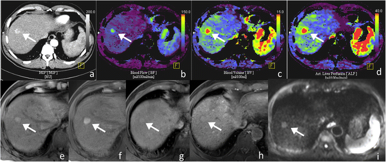
Methods: 36 patients with liver cirrhosis and a total of 67 lesions referred to our hospital for multi-parametric diagnosis of HCC-suspected liver lesions in the setting of liver cirrhosis were prospectively enrolled and underwent PCT and MRI. HCC diagnosis was confirmed either by histology (n = 60) or interval growth (n = 7). For PCT, mean/max blood flow (BF), blood volume (BV), k-trans, arterial liver perfusion (ALP), portal venous perfusion (PVP) and hepatic perfusion index (HPI) were quantified. Two readers identified the lesions based on single maps each being blinded to the number of lesions. MRI-protocol included fat-suppressed T1w-VIBE sequences obtained before, 2, 5, 10 and 20 min after the injection of gadoxetic acid as well as non-enhanced coronal HASTE, axial T1w-VIBE, fat-suppressed T2w-TSE and DWI. Quantitative analysis was performed using enhancement ratios between tumor and liver parenchyma for post-contrast in the hepatobiliary phase (RIRHB), arterial (ERa) and late-venous (ERv) phases as well as signal intensity ratios (liver/parenchyma) on T1w (RIRT1) and T2w (RIRT2).
Results: In PCT analysis, all lesions exhibited high BFmax values (63-250 mL/100 g tissue) and were visible on HPI maps with high degrees of arterial blood supply of (HPI > 96%). In MRI, RIRHB was negative in 8/67. 12/67 HCCs were missed on DWI. 46/67 HCCs showed wash-in and 47/67 HCC showed wash-out of contrast agent. 6/67 HCCs were missed on T1w and 11/67 were missed on T2w-sequences when analyzed separately, while analysis of multiparametric MRI combining typical enhancement pattern, visibility on hepatobiliary phase and T1w-images the same number of lesions as PCT irrespective of their size (1-19 cm) were detected. Quantification of early enhancement by ERa or ERv did not improve detection rates.
Conclusions: Perfusion-CT and gadoxetic acid-enhanced MRI were comparable in detecting HCC lesions. For PCT a mean HPI > 96% proved to be a very robust parameter for detection and characterization of HCC.
Keywords: Hepatocellular Carcinoma; MRI; contrast agent; perfusion; perfusion-CT.
Conflict of interest statement
Ethics approval and consent to participate
All procedures were in accordance with the ethical standards of the institutional and the national research committee and with the Helsinki declaration and its later amendments or comparable ethical standards. Prior to the procedure, all patients gave their informed consent for the procedure. The study was approved by the local Ethics Committee.
Consent for publication
All study subjects gave their written informed consent.
Competing interests
The authors declare that they have no competing interests.
Publisher’s Note
Springer Nature remains neutral with regard to jurisdictional claims in published maps and institutional affiliations.
Figures

Similar articles
-
Differences Between CT-Perfusion and Biphasic Contrast-enhanced CT for Detection and Characterization of Hepatocellular Carcinoma: Potential Explanations for Discrepant Cases.Anticancer Res. 2021 Mar;41(3):1451-1458. doi: 10.21873/anticanres.14903. Anticancer Res. 2021. PMID: 33788737
-
Evaluation of Texture Analysis Parameter for Response Prediction in Patients with Hepatocellular Carcinoma Undergoing Drug-eluting Bead Transarterial Chemoembolization (DEB-TACE) Using Biphasic Contrast-enhanced CT Image Data: Correlation with Liver Perfusion CT.Acad Radiol. 2017 Nov;24(11):1352-1363. doi: 10.1016/j.acra.2017.05.006. Epub 2017 Jun 23. Acad Radiol. 2017. PMID: 28652049
-
Characterization of hepatocellular carcinoma (HCC) lesions using a novel CT-based volume perfusion (VPCT) technique.Eur J Radiol. 2015 Jun;84(6):1029-35. doi: 10.1016/j.ejrad.2015.02.020. Epub 2015 Mar 6. Eur J Radiol. 2015. PMID: 25816994
-
Diagnostic performance of contrast-enhanced multidetector computed tomography and gadoxetic acid disodium-enhanced magnetic resonance imaging in detecting hepatocellular carcinoma: direct comparison and a meta-analysis.Abdom Radiol (NY). 2016 Oct;41(10):1960-72. doi: 10.1007/s00261-016-0807-7. Abdom Radiol (NY). 2016. PMID: 27318936 Free PMC article.
-
Gadoxetic acid-enhanced magnetic resonance imaging: Hepatocellular carcinoma and mimickers.Clin Mol Hepatol. 2019 Sep;25(3):223-233. doi: 10.3350/cmh.2018.0107. Epub 2019 Jan 21. Clin Mol Hepatol. 2019. PMID: 30661336 Free PMC article. Review.
Cited by
-
Role of radiomics in the diagnosis and treatment of gastrointestinal cancer.World J Gastroenterol. 2022 Nov 14;28(42):6002-6016. doi: 10.3748/wjg.v28.i42.6002. World J Gastroenterol. 2022. PMID: 36405385 Free PMC article. Review.
-
Colorectal liver metastases patients prognostic assessment: prospects and limits of radiomics and radiogenomics.Infect Agent Cancer. 2023 Mar 16;18(1):18. doi: 10.1186/s13027-023-00495-x. Infect Agent Cancer. 2023. PMID: 36927442 Free PMC article. Review.
-
Hepatic tumors: pitfall in diagnostic imaging.Acta Biomed. 2020 Jul 13;91(8-S):9-17. doi: 10.23750/abm.v91i8-S.9969. Acta Biomed. 2020. PMID: 32945274 Free PMC article. Review.
-
Radiomics and machine learning analysis by computed tomography and magnetic resonance imaging in colorectal liver metastases prognostic assessment.Radiol Med. 2023 Nov;128(11):1310-1332. doi: 10.1007/s11547-023-01710-w. Epub 2023 Sep 11. Radiol Med. 2023. PMID: 37697033
-
Rim Enhancement after Technically Successful Transarterial Chemoembolization in Hepatocellular Carcinoma: A Potential Mimic of Incomplete Embolization or Reactive Hyperemia?Tomography. 2022 Apr 15;8(2):1148-1158. doi: 10.3390/tomography8020094. Tomography. 2022. PMID: 35448728 Free PMC article.
References
MeSH terms
Substances
LinkOut - more resources
Full Text Sources
Other Literature Sources
Medical
Miscellaneous

