. 2017 Aug 24;91(18):e00838-17.
doi: 10.1128/JVI.00838-17. Print 2017 Sep 15.

Inhibition of Poxvirus Gene Expression and Genome Replication by Bisbenzimide Derivatives

Affiliations

Inhibition of Poxvirus Gene Expression and Genome Replication by Bisbenzimide Derivatives

Artur Yakimovich et al. J Virol. .

Abstract

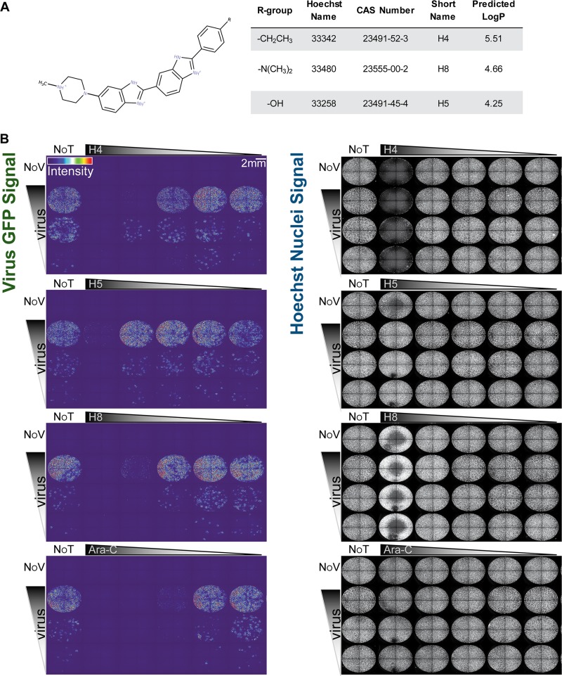
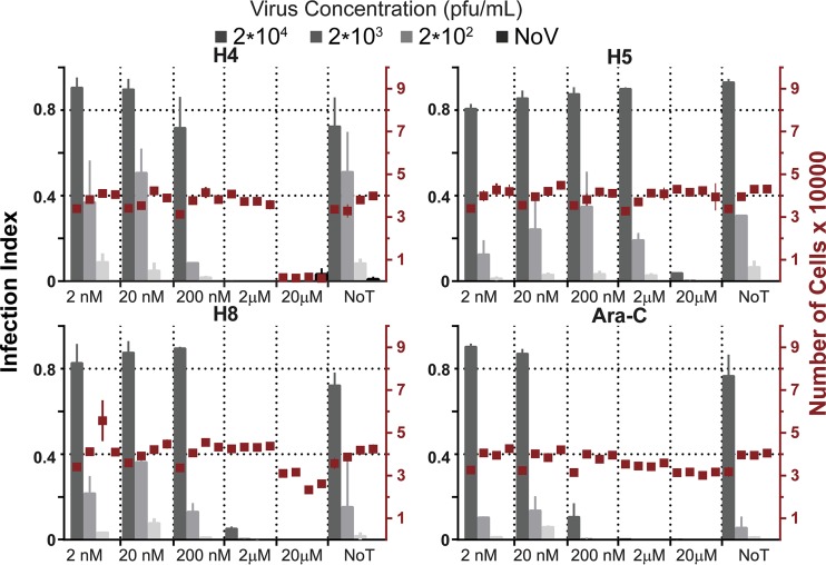
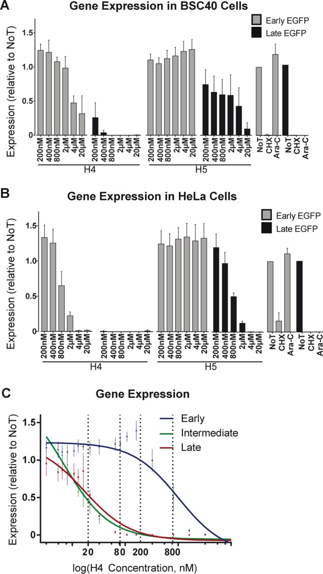
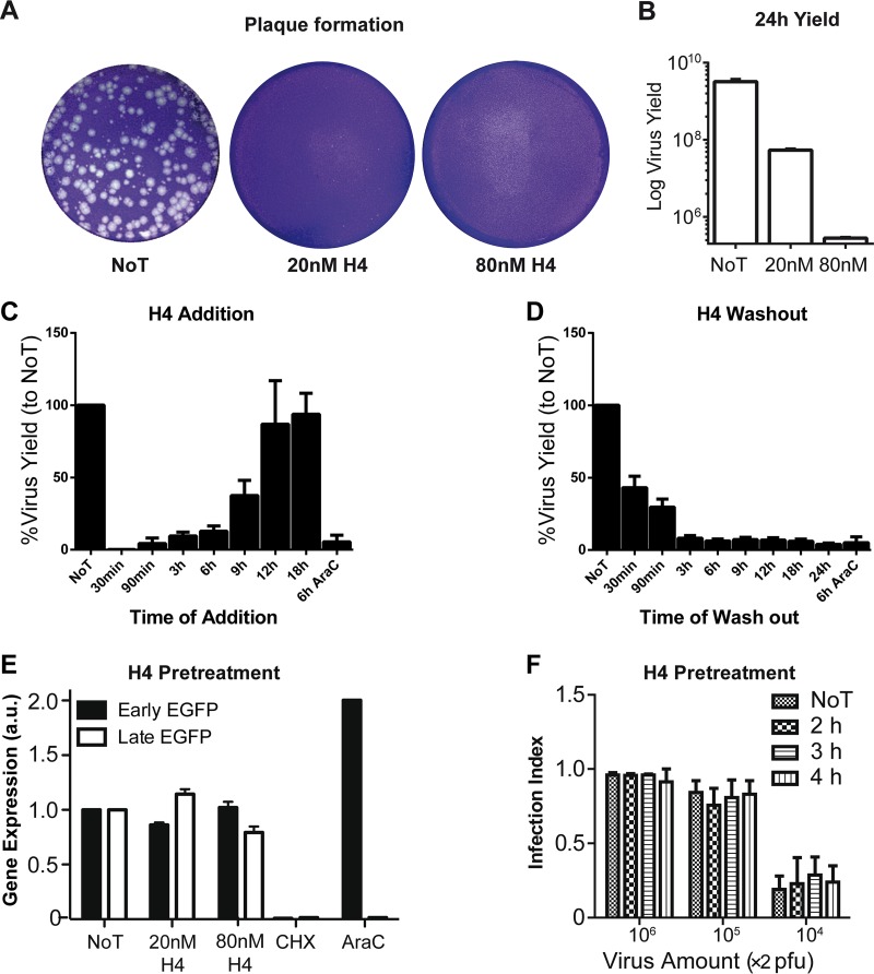
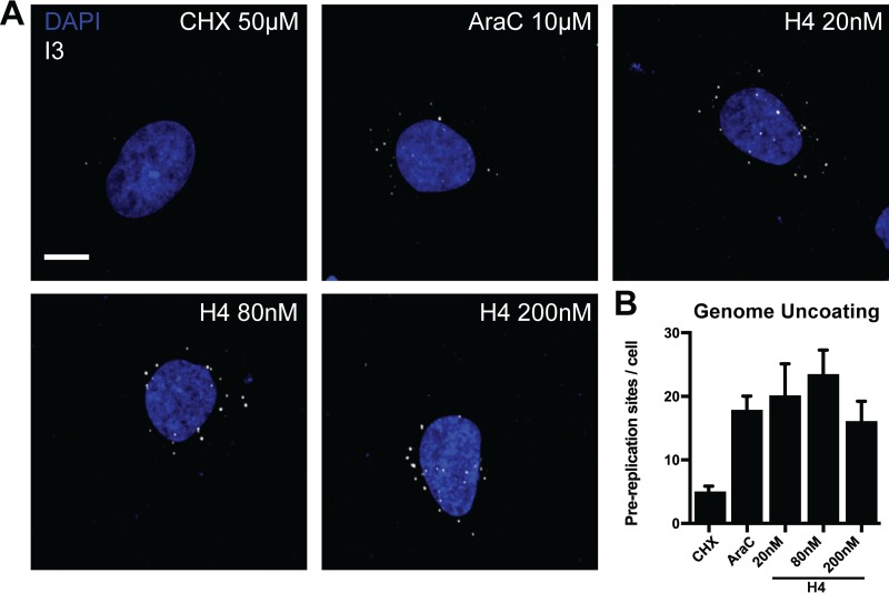
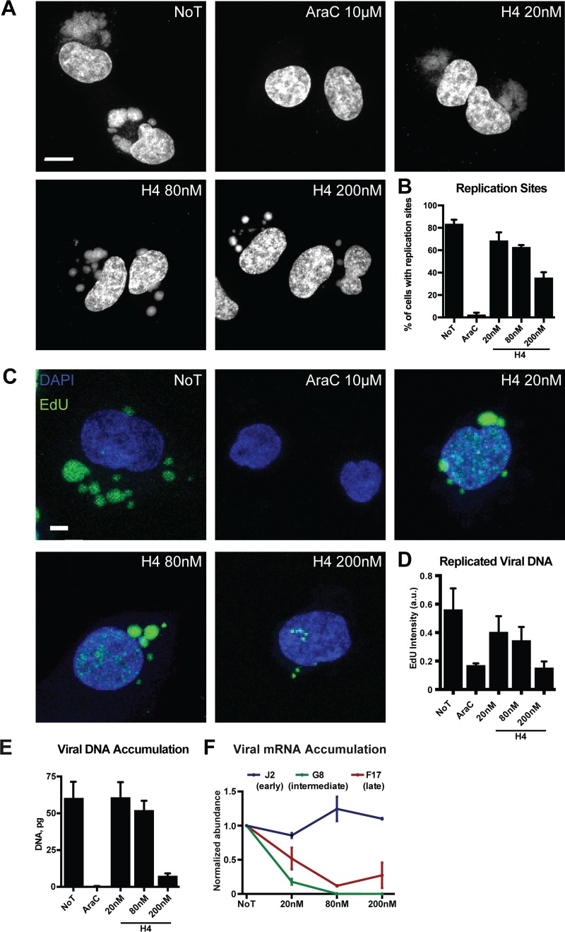
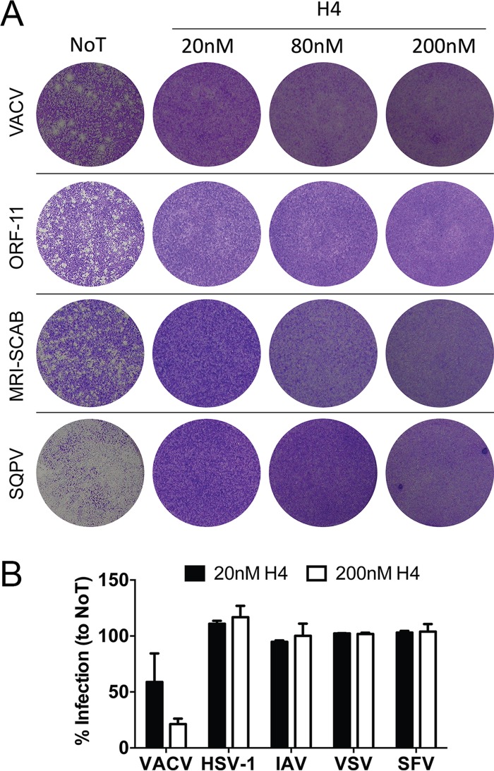
Virus infection of humans and livestock can be devastating for individuals and populations, sometimes resulting in large economic and societal impact. Prevention of virus disease by vaccination or antiviral agents is difficult to achieve. A notable exception was the eradication of human smallpox by vaccination over 30 years ago. Today, humans and animals remain susceptible to poxvirus infections, including zoonotic poxvirus transmission. Here we identified a small molecule, bisbenzimide (bisbenzimidazole), and its derivatives as potent agents against prototypic poxvirus infection in cell culture. We show that bisbenzimide derivatives, which preferentially bind the minor groove of double-stranded DNA, inhibit vaccinia virus infection by blocking viral DNA replication and abrogating postreplicative intermediate and late gene transcription. The bisbenzimide derivatives are potent against vaccinia virus and other poxviruses but ineffective against a range of other DNA and RNA viruses. The bisbenzimide derivatives are the first inhibitors of their class, which appear to directly target the viral genome without affecting cell viability.IMPORTANCE Smallpox was one of the most devastating diseases in human history until it was eradicated by a worldwide vaccination campaign. Due to discontinuation of routine vaccination more than 30 years ago, the majority of today's human population remains susceptible to infection with poxviruses. Here we present a family of bisbenzimide (bisbenzimidazole) derivatives, known as Hoechst nuclear stains, with high potency against poxvirus infection. Results from a variety of assays used to dissect the poxvirus life cycle demonstrate that bisbenzimides inhibit viral gene expression and genome replication. These findings can lead to the development of novel antiviral drugs that target viral genomes and block viral replication.

Keywords: DNA replication; antiviral agents; poxvirus; vaccinia virus; viral transcription.


Figures

FIG 1
FIG 1
(A) Chemical structure, properties including partitioning coefficient (LogP), and compound information of the bisbenzimides used in this study. (B) Bisbenzimides (H4, H5, and H8) block VACV replication in tissue culture. BSC40 cells were infected with a serial dilution of E/L EGFP VACV and treated with serial dilutions of H4, H5, H8, or AraC. Full-well images show EGFP-expressing infected cells color coded by intensity (left panels). Nuclei were detected by staining with Hoechst (right panels). Experiments were performed in triplicate; representative images are displayed. NoT, not drug treated; NoV, not virus infected.
FIG 2
FIG 2
Quantification of infected EGFP-expressing cells and total cell number (nuclei) from Fig. 1B. Gray bars indicate the infection index, and red boxes indicate the cell number for each condition tested. Experiments were performed in triplicate. Results are displayed as means ± standard deviations (SD).
FIG 3
FIG 3
Bisbenzimides inhibit VACV intermediate and late gene expression. (A and B) BSC40 (A) or HeLa (B) cells were infected with WR E EGFP (gray bars) or WR L EGFP (black bars) VACV. Cells were scored for EGFP expression by flow cytometry, and infected cells were quantified relative to untreated cells. CHX and AraC served as controls for these experiments. (C) HeLa cells treated with various concentrations of H4 were infected with WR E EGFP (blue line), WR I EGFP (green line), or WR L EGFP (red line), and the percentage of EGFP-positive infected cells was quantified by flow cytometry. These values were fitted to dose-response curves to estimate EC50 and EC90 values (dashed lines).
FIG 4
FIG 4
H4 inhibits plaque formation, reduces virus yield, and blocks early VACV infection without impacting particle infectivity. (A) HeLa cells were infected with 150 PFU of WT VACV, and infection was allowed to proceed for 72 h. The plates were stained with crystal violet to visualize plaques. (B) HeLa cells were infected with WT VACV (MOI of 1). Twenty-four hours postinfection, cells were harvested and lysed and virus yield was determined by titration and plaque formation. (C) HeLa cells were infected with WT virus (MOI of 1), and 80 nM H4 was added at the indicated time points. A sample subjected to AraC addition at 6 hpi was included as a positive control for inhibition. Cells were harvested 24 hpi, and virus yield was determined for each sample by serial dilution plaque assay. (D) HeLa cells were infected with WT virus (MOI of 1) in the presence of 80 nM H4. At the indicated time points, cells were washed, and infection was allowed to proceed. A sample subjected to AraC washout at 6 hpi was included as a positive control for inhibition. At 24 hpi, virus yield was determined for each sample by serial dilution plaque assay. (E) WR E EGFP (black bars) or WR L EGFP (white bars) virions were preincubated with 20 or 80 nM H4 for 30 min at room temperature. Virus particles were washed three times and used to infect HeLa cells. Samples were analyzed by flow cytometry for infected EGFP-positive cells at 6 hpi (early) and 8 hpi (late). a.u., absorbance units. (F) WR E/L EGFP virions were preincubated with 2 μM H4 for 2, 3, or 4 h. Virions were washed and used to infect HeLa cells prior to fixation and analysis by Plaque 2.0 for total nuclei and EGFP-positive infected cells. (A to F) All experiments were performed in triplicate; representative images are shown (A), or results are displayed as means ± SD (B to F).
FIG 5
FIG 5
H4 does not impact viral genome uncoating. (A) HeLa cells were infected with WT VACV (MOI of 10) in the presence of 20, 80, or 200 nM H4 and AraC. Prereplication sites were visualized by immunofluorescence staining against I3 followed by confocal microscopy. CHX and AraC served as controls for uncoating and replication, respectively. (B) Quantification of prereplication sites per cell from panel A. (A and B) Experiments were performed in triplicate; representative images are displayed, and results are shown as means ± standard errors of the means (SEM). Scale bar, 10 μm.
FIG 6
FIG 6
H4 attenuates VACV IG/LG transcription and DNA replication in a dose-dependent fashion. (A) HeLa cells were infected (MOI of 10) in the presence of 20, 80, or 200 nM H4. At 8 hpi, cells were fixed and stained with DAPI and imaged by confocal microscopy. Scale bar, 10 μm. (B) The number of cells with cytoplasmic replication sites was quantified per condition. AraC served as a control for inhibition of DNA replication site formation. (C) HeLa cells were infected (MOI of 10) in the presence of 20, 80, or 200 nM H4 and EdU. At 8 hpi, EdU incorporation was detected with a Click-iT EdU imaging kit followed by confocal microscopy. Scale bar, 10 μm. (D) The total intensity of EdU incorporation into replication sites was quantified. (E) The amount of viral DNA from cells infected in the absence or presence of H4 at different concentrations was quantified by qPCR at 8 hpi. AraC served as a control for inhibition of DNA replication. (F) The levels of early (J2), intermediate (G8), and late (F17) viral mRNA from infected HeLa cells were quantified by RT-qPCR. Cells were infected in the absence or presence of various concentrations of H4, and RT was performed at 2 hpi for J2, 4 hpi for G8, and 8 hpi for F17. Results are displayed as the average abundance normalized to untreated samples. All experiments were performed in triplicate; representative images (A and C) or means ± SD (B and D to F) are displayed.
FIG 7
FIG 7
Poxviruses are acutely sensitive to H4 inhibitory activity. (A) Fetal lamb skin cell monolayers were infected with VACV or ORF-11 (100 PFU), MRI-SCAB (500 PFU), or squirrelpox virus (SQPV [1,000 PFU]). Cells were fixed and plaques visualized by crystal violet staining at 3 days (VACV), 4 days (ORF-11), or 7 days (MRI-SCAB and SQPV). Experiments were performed in triplicate; representative images are shown. (B) HeLa cells were infected with EGFP-expressing variants of VACV WR, herpes simplex virus 1 (HSV-1), influenza A virus (IAV), vesicular stomatitis virus (VSV), or Semliki Forest virus (SFV). For each, infection was allowed to proceed for 6 to 8 h, after which cells were analyzed for EGFP expression by flow cytometry. Experiments were performed in triplicate; the percentage of infection relative to untreated (NoT) controls is displayed as the mean ± SD.

Similar articles

Cited by

References

    1. Lederberg J. 2000. Infectious history. Science 288:287–293. doi:10.1126/science.288.5464.287. - DOI - PubMed
    1. Greber UF, Bartenschlager R. 2017. Editorial: an expanded view of viruses. FEMS Microbiol Rev 41:1–4. doi:10.1093/femsre/fuw044. - DOI - PMC - PubMed
    1. Koonin EV, Dolja VV, Krupovic M. 2015. Origins and evolution of viruses of eukaryotes: the ultimate modularity. Virology 479:2–25. doi:10.1016/j.virol.2015.02.039. - DOI - PMC - PubMed
    1. Prasad V, Suomalainen M, Hemmi S, Greber UF. 2017. The cell cycle-dependent kinase Cdk9 is a postexposure drug target against human adenoviruses. ACS Infect Dis 3:398–405. doi:10.1021/acsinfecdis.7b00009. - DOI - PubMed
    1. Santos-Fernandes E, Beltrame CO, Byrd CM, Cardwell KB, Schnellrath LC, Medaglia ML, Hruby DE, Jordan R, Damaso CR. 2013. Increased susceptibility of Cantagalo virus to the antiviral effect of ST-246. Antiviral Res 97:301–311. doi:10.1016/j.antiviral.2012.11.010. - DOI - PubMed

Publication types

LinkOut - more resources