

Linoleic acid participates in the response to ischemic brain injury through oxidized metabolites that regulate neurotransmission
- PMID: 28659576
- PMCID: PMC5489485
- DOI: 10.1038/s41598-017-02914-7
Linoleic acid participates in the response to ischemic brain injury through oxidized metabolites that regulate neurotransmission
Abstract
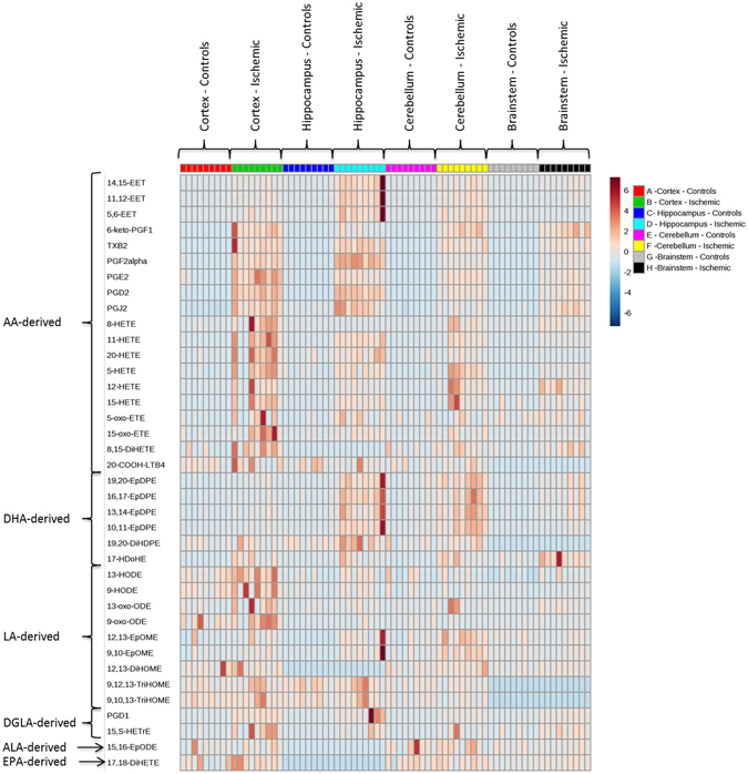
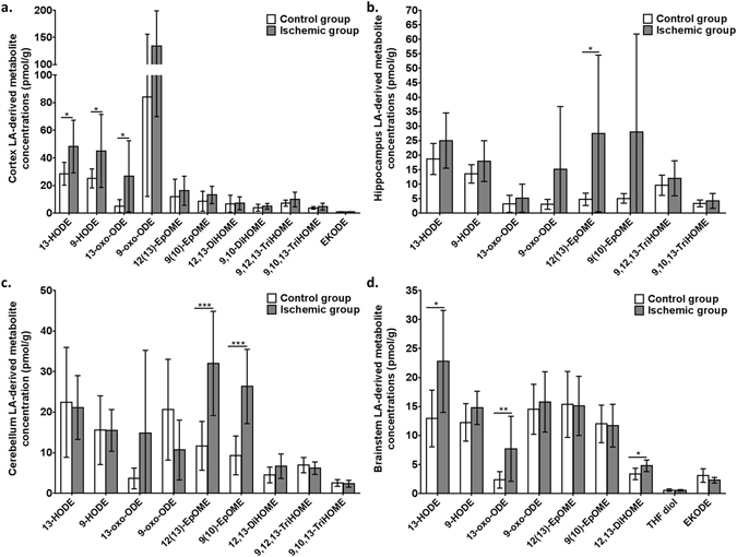
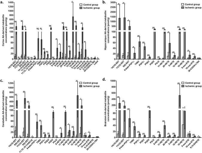
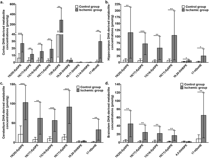
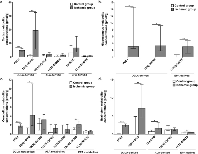
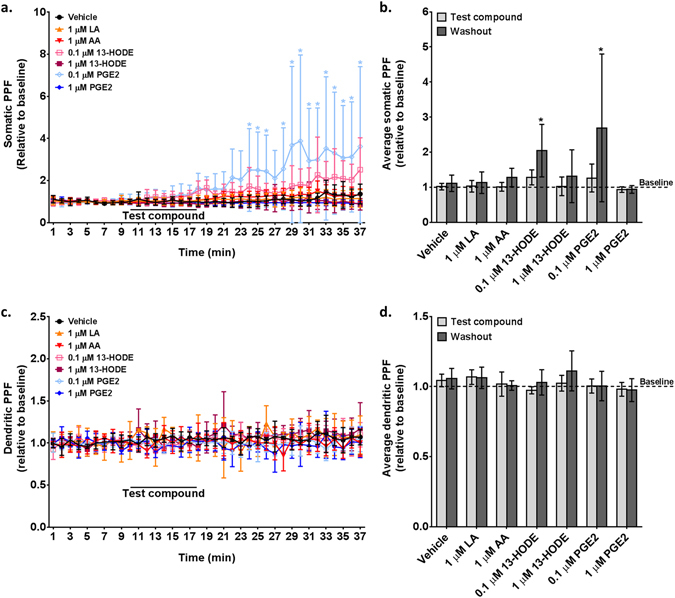
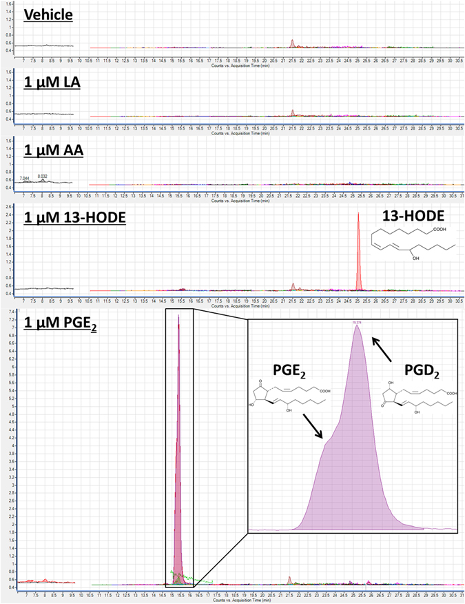
Linoleic acid (LA; 18:2 n-6), the most abundant polyunsaturated fatty acid in the US diet, is a precursor to oxidized metabolites that have unknown roles in the brain. Here, we show that oxidized LA-derived metabolites accumulate in several rat brain regions during CO2-induced ischemia and that LA-derived 13-hydroxyoctadecadienoic acid, but not LA, increase somatic paired-pulse facilitation in rat hippocampus by 80%, suggesting bioactivity. This study provides new evidence that LA participates in the response to ischemia-induced brain injury through oxidized metabolites that regulate neurotransmission. Targeting this pathway may be therapeutically relevant for ischemia-related conditions such as stroke.
Conflict of interest statement
The authors declare that they have no competing interests.
Figures
Similar articles
-
Linoleic acid-derived metabolites constitute the majority of oxylipins in the rat pup brain and stimulate axonal growth in primary rat cortical neuron-glia co-cultures in a sex-dependent manner.J Neurochem. 2020 Jan;152(2):195-207. doi: 10.1111/jnc.14818. Epub 2019 Nov 26. J Neurochem. 2020. PMID: 31283837 Free PMC article.
-
Effects of diets enriched in linoleic acid and its peroxidation products on brain fatty acids, oxylipins, and aldehydes in mice.Biochim Biophys Acta Mol Cell Biol Lipids. 2018 Oct;1863(10):1206-1213. doi: 10.1016/j.bbalip.2018.07.007. Epub 2018 Jul 25. Biochim Biophys Acta Mol Cell Biol Lipids. 2018. PMID: 30053599 Free PMC article.
-
Feeding mice a diet high in oxidized linoleic acid metabolites does not alter liver oxylipin concentrations.Prostaglandins Leukot Essent Fatty Acids. 2021 Sep;172:102316. doi: 10.1016/j.plefa.2021.102316. Epub 2021 Jun 24. Prostaglandins Leukot Essent Fatty Acids. 2021. PMID: 34403987 Free PMC article.
-
Hydroxyoctadecadienoic acids: Oxidised derivatives of linoleic acid and their role in inflammation associated with metabolic syndrome and cancer.Eur J Pharmacol. 2016 Aug 15;785:70-76. doi: 10.1016/j.ejphar.2015.03.096. Epub 2015 May 15. Eur J Pharmacol. 2016. PMID: 25987423 Review.
-
Dietary factors and growth and metabolism in experimental tumors.J Nutr Biochem. 2007 Oct;18(10):637-49. doi: 10.1016/j.jnutbio.2006.12.009. Epub 2007 Apr 5. J Nutr Biochem. 2007. PMID: 17418560 Review.
Cited by
-
Acute Hypercapnia/Ischemia Alters the Esterification of Arachidonic Acid and Docosahexaenoic Acid Epoxide Metabolites in Rat Brain Neutral Lipids.Lipids. 2020 Jan;55(1):7-22. doi: 10.1002/lipd.12197. Epub 2019 Nov 6. Lipids. 2020. PMID: 31691988 Free PMC article.
-
Soluble Epoxide Hydrolase-Derived Linoleic Acid Oxylipins in Serum Are Associated with Periventricular White Matter Hyperintensities and Vascular Cognitive Impairment.Transl Stroke Res. 2019 Oct;10(5):522-533. doi: 10.1007/s12975-018-0672-5. Epub 2018 Nov 15. Transl Stroke Res. 2019. PMID: 30443886
-
Linoleic acid-good or bad for the brain?NPJ Sci Food. 2020 Jan 2;4:1. doi: 10.1038/s41538-019-0061-9. eCollection 2020. NPJ Sci Food. 2020. PMID: 31909187 Free PMC article. Review.
-
Repeated Low-Level Blast Exposure Alters Urinary and Serum Metabolites.Metabolites. 2023 May 8;13(5):638. doi: 10.3390/metabo13050638. Metabolites. 2023. PMID: 37233679 Free PMC article.
-
Spatial quantitative metabolomics enables identification of remote and sustained ipsilateral cortical metabolic reprogramming after stroke.Nat Metab. 2025 Aug 5. doi: 10.1038/s42255-025-01340-8. Online ahead of print. Nat Metab. 2025. PMID: 40764673
References
-
- Ellis EF, Wei EP, Kontos HA. Vasodilation of cat cerebral arterioles by prostaglandins D2, E2, G2, and I2. Am J Physiol. 1979;237:H381–385. - PubMed
Publication types
MeSH terms
Substances
Grants and funding
LinkOut - more resources
Full Text Sources
Other Literature Sources
