. 2017:2017:8248175.
doi: 10.1155/2017/8248175. Epub 2017 Jun 4.

Type II Endometrial Cancer Overexpresses NILCO: A Preliminary Evaluation

Affiliations

Type II Endometrial Cancer Overexpresses NILCO: A Preliminary Evaluation

Danielle Daley-Brown et al. Dis Markers. 2017.

Abstract

Objective: The expression of NILCO molecules (Notch, IL-1, and leptin crosstalk outcome) and the association with obesity were investigated in types I and II endometrial cancer (EmCa). Additionally, the involvement of NILCO in leptin-induced invasiveness of EmCa cells was investigated.

Methods: The expression of NILCO mRNAs and proteins were analyzed in EmCa from African-American (n = 29) and Chinese patients (tissue array, n = 120 cases). The role of NILCO in leptin-induced invasion of Ishikawa and An3ca EmCa cells was investigated using Notch, IL-1, and leptin signaling inhibitors.

Results: NILCO molecules were expressed higher in type II EmCa, regardless of ethnic background or obesity status of patients. NILCO proteins were mainly localized in the cellular membrane and cytoplasm of type II EmCa. Additionally, EmCa from obese African-American patients showed higher levels of NILCO molecules than EmCa from lean patients. Notably, leptin-induced EmCa cell invasion was abrogated by NILCO inhibitors.

Conclusion: Type II EmCa expressed higher NILCO molecules, which may suggest it is involved in the progression of the more aggressive EmCa phenotype. Obesity was associated with higher expression of NILCO molecules in EmCa. Leptin-induced cell invasion was dependent on NILCO. Hence, NILCO might be involved in tumor progression and could represent a new target/biomarker for type II EmCa.


Figures
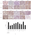
Figure 1
Figure 1
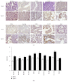
Immunohistochemical (IHC) detection of NILCO and targets in EmCa from African-American women. (a) Representative IHC pictures from type I (n = 7; 41%) and type II (n = 10; 59%) EmCa from obese African-American women. (b) Histogram of the semiquantitative HSCORE values. Brown color characterizes positive staining. Magnification ×40. p < 0.05.
Figure 2
Figure 2
HSCORE values from lean and obese African-American women. (a) HSCORE of lean type I (n = 5; 42%) versus lean type II (n = 7; 58%). (b) HSCORE of lean type I (n = 5; 42%) versus obese type I (n = 7; 41%). (c) HSCORE of lean type II (n = 7; 58%) versus obese type II (n = 10; 59%). The expression levels of NILCO components and targets were analyzed in EmCa from lean (n = 12) versus obese (n = 17) women. p < 0.05.
Figure 3
Figure 3
Immunohistochemical (IHC) detection of NILCO and targets in EmCa tissue microarrays from Chinese women. (a) Representative IHC pictures from type I (n = 97; 81%) and type II (n = 23; 19%) EmCa from Chinese women. (b) Histogram of the semiquantitative HSCORE values. Brown color characterizes positive staining. Magnification ×40. p < 0.05.
Figure 4
Figure 4
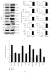
Protein and mRNA expression of Notch receptors, ligands, and molecular targets in type I and type II EmCa from African-American women. (a) Western blot representative data of NILCO components in African-American women. Type I (n = 8); type II (n = 6). (b) mRNA expression levels of NILCO and targets in African-American women by real-time PCR. mRNA levels were normalized to GAPDH. p < 0.05.
Figure 5
Figure 5
Leptin and IL-1β levels in EmCa lysates. (a) Leptin levels in EmCa. (b) IL-1β levels in EmCa. Type I EmCa (n = 8) and type II EmCa (n = 6) lysates were analyzed by ELISA (R&D systems).
Figure 6
Figure 6
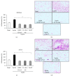
Leptin-induced EmCa cell invasion was abrogated by inhibition of Notch and IL-1 signaling. (a) Quantitative assessment of Ishikawa (derived from type I EmCa) cell invasion. (b) Representative pictures of the effects of leptin and NILCO inhibitors on Ishikawa cell invasion. (c) Quantitative assessment of An3ca (derived from type II EmCa) cell invasion. (d) Representative pictures of the effects of leptin and NILCO inhibitors on An3ca cell invasion. Results from cell migration (Boyden chamber cell invasion assay; Corning BioCoat Matrigel invasion chamber) were obtained after 24 h and normalized to basal conditions. Invading cells were detected by hematoxylin staining (see Section 2). Data (mean ± standard error) representative results derived from a minimum of 3 independent experiments. p < 0.05.

References

    1. Daley-Brown D., Oprea-Ilies G. M., Lee R., Pattillo R., Gonzalez-Perez R. R. Molecular cues on obesity signals, tumor markers and endometrial cancer. Hormone Molecular Biology and Clinical Investigation. 2015;21(1):89–106. doi: 10.1515/hmbci-2014-0049. - DOI - PMC - PubMed
    1. Siegel R. L., Miller K. D., Jemal A. Cancer statistics, 2015. CA Cancer Journal for Clinicians. 2014;65(1):5–29. doi: 10.3322/caac.21254. - DOI - PubMed
    1. Collins Y., Holcomb K., Chapman-Davis E., Khabele D., Farley J. H. Gynecologic cancer disparities: a report from the health disparities taskforce of the Society of Gynecologic Oncology. Gynecologic Oncology. 2014;133(3):353–361. doi: 10.1016/j.ygyno.2013.12.039. - DOI - PMC - PubMed
    1. Setiawan V. W., Yang H. P., Pike M. C., et al. Type I and II endometrial cancers: have they different risk factors? Journal of Clinical Oncology. 2013;31(20):2607–2618. doi: 10.1200/JCO.2012.48.2596. - DOI - PMC - PubMed
    1. Barakat R. R., Markman M., Randall M. Corpus: epithelial tumors. Principles and Practice of Gynecologic Oncology. 2009:683–686.

MeSH terms

LinkOut - more resources