. 2017 Jun 12:11:166.
doi: 10.3389/fncel.2017.00166. eCollection 2017.

NMDA Receptors Regulate the Structural Plasticity of Spines and Axonal Boutons in Hippocampal Interneurons

Affiliations

NMDA Receptors Regulate the Structural Plasticity of Spines and Axonal Boutons in Hippocampal Interneurons

Marta Perez-Rando et al. Front Cell Neurosci. .

Abstract

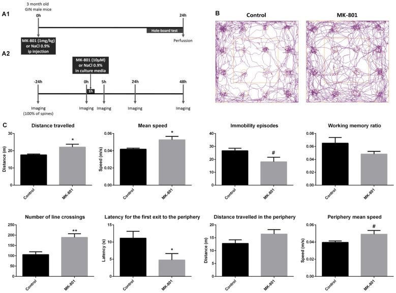
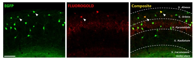
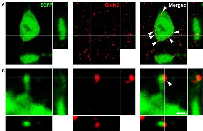
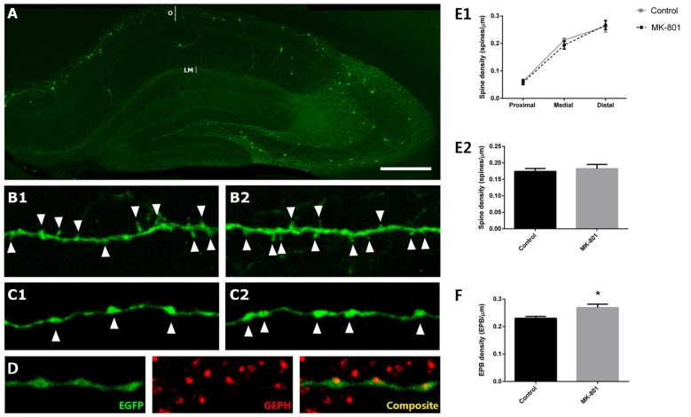
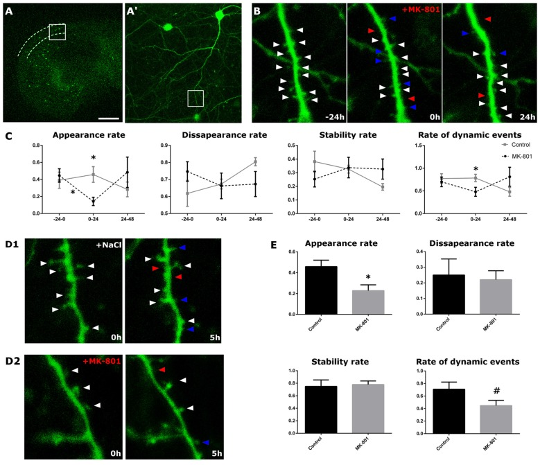
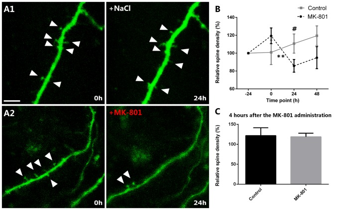
N-methyl-D-aspartate receptors (NMDARs) are present in both pyramidal neurons and interneurons of the hippocampus. These receptors play an important role in the adult structural plasticity of excitatory neurons, but their impact on the remodeling of interneurons is unknown. Among hippocampal interneurons, somatostatin-expressing cells located in the stratum oriens are of special interest because of their functional importance and structural characteristics: they display dendritic spines, which change density in response to different stimuli. In order to understand the role of NMDARs on the structural plasticity of these interneurons, we have injected acutely MK-801, an NMDAR antagonist, to adult mice which constitutively express enhanced green fluorescent protein (EGFP) in these cells. We have behaviorally tested the animals, confirming effects of the drug on locomotion and anxiety-related behaviors. NMDARs were expressed in the somata and dendritic spines of somatostatin-expressing interneurons. Twenty-four hours after the injection, the density of spines did not vary, but we found a significant increase in the density of their en passant boutons (EPB). We have also used entorhino-hippocampal organotypic cultures to study these interneurons in real-time. There was a rapid decrease in the apparition rate of spines after MK-801 administration, which persisted for 24 h and returned to basal levels afterwards. A similar reversible decrease was detected in spine density. Our results show that both spines and axons of interneurons can undergo remodeling and highlight NMDARs as regulators of this plasticity. These results are specially relevant given the importance of all these players on hippocampal physiology and the etiopathology of certain psychiatric disorders.

Keywords: MK-801; NMDAR; axonal boutons; interneurons; organotypic cultures; spine dynamics.


Figures

Figure 1
Figure 1
(A1) Experimental design of the in vivo experiment. (A2) Experimental design of the real-time in vitro experiment. (B,C) Hole-board behavioral test. (B) Representative tracking-plots of NaCl-treated animals (control) and MK-801-treated animals (MK-801). (C) Graphs representing the changes observed in the behavioral parameters that were analyzed (all graphs represent mean + SEM., *p < 0.05, **p < 0.01, #0.05 < p < 0.1).
Figure 2
Figure 2
Characterization of enhanced greenfluorescent protein (EGFP)-expressing interneurons in the stratum oriens of CA1. EGFP+ cells (left), fluorogold retrograde tracing from the medial septal nucleus (middle) and the composite image. Scale bar: 15 μm.
Figure 3
Figure 3
Immunohistochemistry against the N-methyl-D-aspartate receptor (NMDAR) obligatory subunit GluN1 in GIN mice. (A) Puncta expressing NMDARs (arrowheads) in an EGFP labeled soma. (B) Dendritic spine of an EFGP-labeled interneuron. Note the expression of NMDAR on the spine head. Panels on the right and bottom are orthogonal projections of the middle panel. Scale bar: 2 μm in (A), 0.50 μm in (B).
Figure 4
Figure 4
Structural modifications in EGFP-expressing interneurons in the stratum oriens after MK-801 injection. (A) Hippocampus of a GIN mouse. The strata oriens (O) and lacunosum moleculare (LM) are indicated with dotted lines. (B) Segments of dendrites bearing spines (arrowheads) from a control animal (B1) and a MK-801-treated mouse (B2). (C) Segments of axons of somatostatin-expressing interneurons in the stratum lacunosum moleculare showing en passant boutons (EPBs, arrowheads) in control (C1) and MK-801-treated (C2) animals. (D) Axonal segment with three EPB in juxtaposition to gephyrin-expressing puncta (GEPH). (E1) Graph representing the spine density in the proximal, medial and distal dendritic segment relative to the soma. (E2) Graph representing the spine density in the total length of the dendrite. (F) Graph showing the density of EPBs in stratum lacunosum moleculare, where a significant increase can be observed in the MK-801 group (all graphs represent mean + SEM., *p < 0.05). Scale bar: 180 μm in (A), 7 μm in (B), 5 μm in (C) and 7 μm in (D).
Figure 5
Figure 5
Structural dynamics of EGFP-expressing interneurons after MK-801 administration. (A) Panoramic view of an organotypic entorhino-hippocampal culture, with the stratum oriens of CA1 delineated by dotted lines. (A′) Enlarged view of the squared region with an interneuron in the stratum oriens. (B) Enlarged view of the squared region in (A,A′). The figure shows a dendritic segment 24 h prior (left panel), right before (middle panel) and 24 h after (right panel) the MK-801 administration. Arrowheads point to stable (white), gained (blue) and lost (red) spines. (C) Graphs showing different dynamic rates in control and MK-801 treated slices (appearance, disappearance, stability rate and rate of total dynamic events). (D1,D2) Microphotographs showing the differences in the spine appearance rate between a control slice (D1) and the MK-801 treated slice (D2) 5 h after the starting of the MK-801 administration. (E) Graphs showing the different rates 5 h after the beginning of the treatment (all graphs represent mean + SEM., *p < 0.05, #0.05 < p < 0.1). Scale bar: 1500 μm in (A), 150 μm in (A′), 1.5 μm in (B,D).
Figure 6
Figure 6
Relative spine density after MK-801 administration. (A) Representative images of dendritic segments in control (A1) and MK-801-treated (A2) groups. Note the decreased relative spine density 24 h after the MK-801 treatment (A2, right panel). Triangles mark the dendritic spines. (B) Graph showing the relative spine densities in the different time points. (C) Graph representing the relative spine density 5 h after the beginning of the MK-801 administration. Scale bar: 6 μm (All graphs represent mean + SEM., **p < 0.01, #0.05 < p < 0.1).

References

    1. Acsády L., Kamondi A., Sík A., Freund T., Buzsáki G. (1998). GABAergic cells are the major postsynaptic targets of mossy fibers in the rat hippocampus. J. Neurosci. 18, 3386–3403. - PMC - PubMed
    1. Adell A., Jiménez-Sánchez L., López-Gil X., Romón T. (2012). Is the acute NMDA receptor hypofunction a valid model of schizophrenia? Schizophr. Bull. 38, 9–14. 10.1093/schbul/sbr133 - DOI - PMC - PubMed
    1. Alvarez V. A., Ridenour D. A., Sabatini B. L. (2007). Distinct structural and ionotropic roles of NMDA receptors in controlling spine and synapse stability. J. Neurosci. 27, 7365–7376. 10.1523/jneurosci.0956-07.2007 - DOI - PMC - PubMed
    1. Bailey C. H., Bartsch D., Kandel E. R. (1996). Toward a molecular definition of long-term memory storage. Proc. Natl. Acad. Sci. U S A 93, 13445–13452. 10.1073/pnas.93.24.13445 - DOI - PMC - PubMed
    1. Becker N., Wierenga C. J., Fonseca R., Bonhoeffer T., Nägerl U. V. (2008). LTD induction causes morphological changes of presynaptic boutons and reduces their contacts with spines. Neuron 60, 590–597. 10.1016/j.neuron.2008.09.018 - DOI - PubMed