. 2017 Jun 13:8:395.
doi: 10.3389/fphys.2017.00395. eCollection 2017.

Cobalt Chloride Upregulates Impaired HIF-1α Expression to Restore Sevoflurane Post-conditioning-Dependent Myocardial Protection in Diabetic Rats

Affiliations

Cobalt Chloride Upregulates Impaired HIF-1α Expression to Restore Sevoflurane Post-conditioning-Dependent Myocardial Protection in Diabetic Rats

Jianjiang Wu et al. Front Physiol. .

Abstract

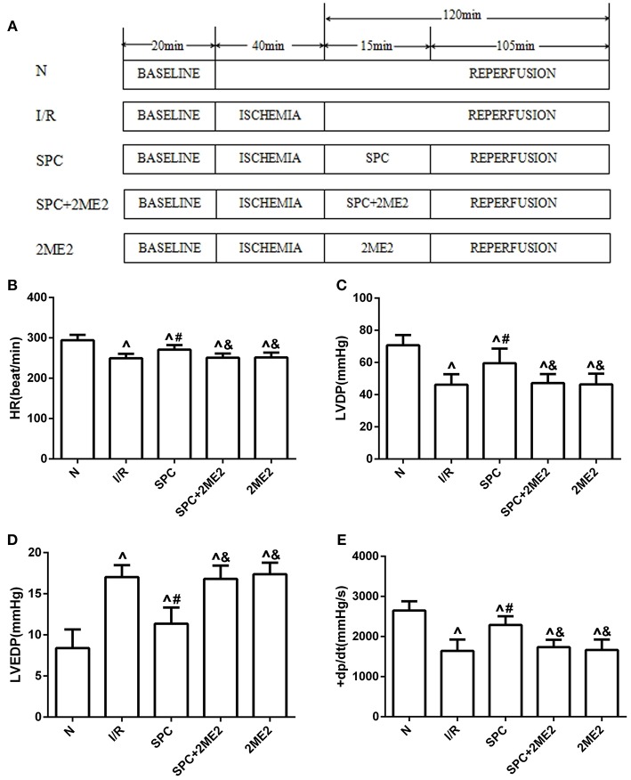
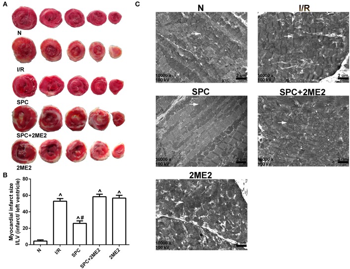
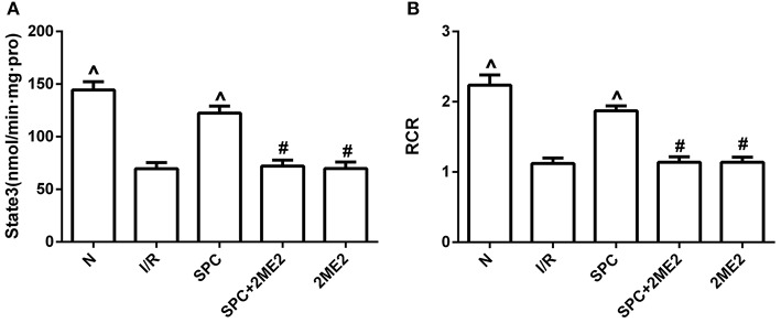
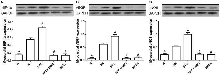
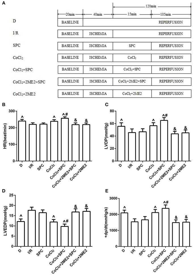
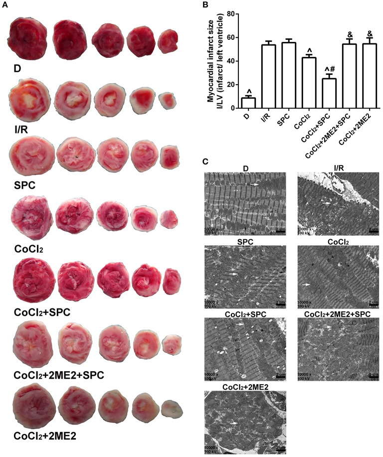
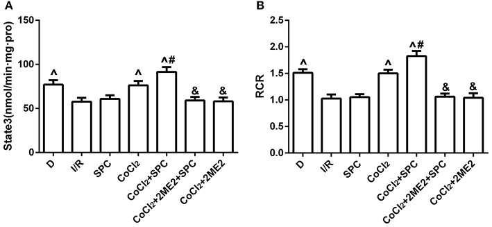
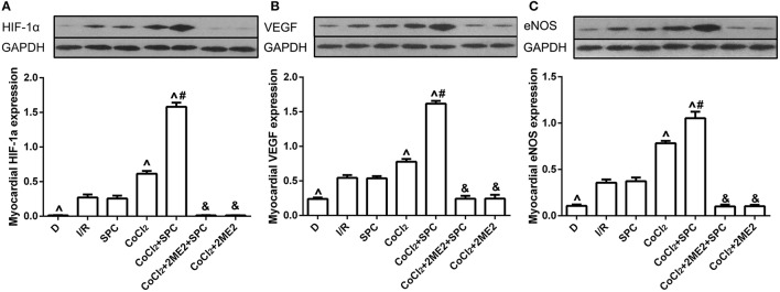
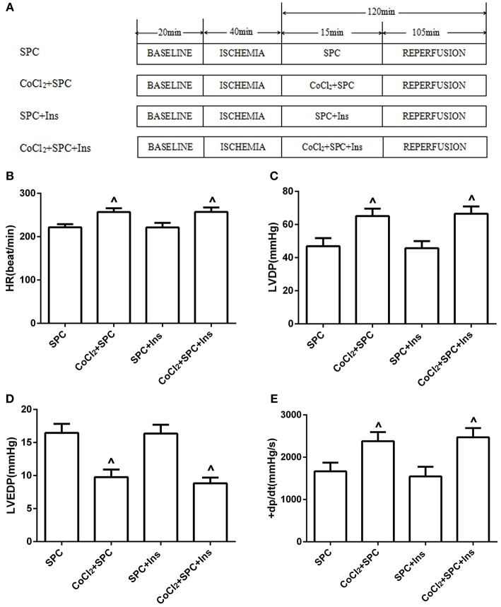
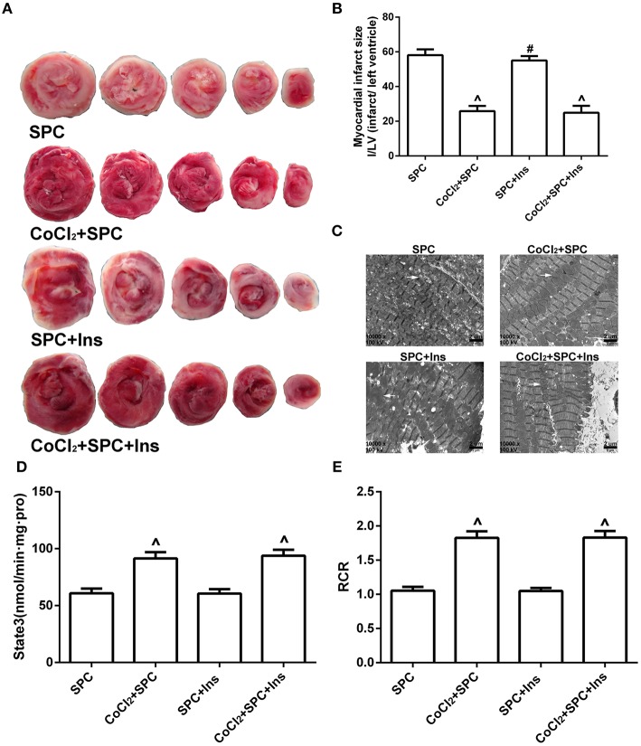
Previous studies from our group have demonstrated that sevoflurane post-conditioning (SPC) protects against myocardial ischemia reperfusion injury via elevating the intranuclear expression of hypoxia inducible factor-1 alpha (HIF-1α). However, diabetic SPC is associated with decreased myocardial protection and disruption of the HIF-1 signaling pathway. Previous studies have demonstrated that cobalt chloride (CoCl2) can upregulate HIF-1α expression under diabetic conditions, but whether myocardial protection by SPC can be restored afterward remains unclear. We established a rat model of type 2 diabetes and a Langendorff isolated heart model of ischemia-reperfusion injury. Prior to reperfusion, 2.4% sevoflurane was used as a post-conditioning treatment. The diabetic rats were treated with CoCl2 24 h before the experiment. At the end of reperfusion, tests were performed to assess myocardial function, infarct size, mitochondrial morphology, nitric oxide (NO), Mitochondrial reactive oxygen species (ROS), mitochondrial respiratory function and enzyme activity, HIF-1α, vascular endothelial growth factor (VEGF) and endothelial NO synthase (eNOS) protein levels. In addition, myocardial protection by SPC was monitored after the blood glucose levels were lowered by insulin. The diabetic state was associated with deficient SPC protection and decreased HIF-1α expression. After treating the diabetic rats with CoCl2, SPC significantly upregulated the expression of HIF-1α, VEGF and eNOS, which markedly improved cardiac function, NO, mitochondrial respiratory function, and enzyme activity and decreased the infarction areas and ROS. In addition, these effects were not influenced by blood glucose levels. This study proved that CoCl2activates the HIF-1α signaling pathway, which restores SPC-dependent myocardial protection under diabetic conditions, and the protective effects of SPC were independent of blood glucose levels.

Keywords: diabetic state; hypoxia inducible factor-1; ischemia-reperfusion; myocardial protection; sevoflurane post-conditioning.


Figures

Figure 1
Figure 1
The schematic diagram of the experimental procedures. Except the N group, all hearts were subjected to 40 min whole heart ischemia, followed by reperfusion for 120 min. The SPC received 2.4% sevoflurane (1.0 MAC) treatment for 15 min, followed by reperfusion for 105 min. (A) The schematic diagram of non-diabetic rats. The cardiac function indicators under non-diabetic state. (B) Heart rate (HR, beat/per min). (C) Left ventricular developed pressure (LVDP, mmHg). (D) Left ventricular end-diastolic pressure (LVEDP, mmHg). (E) Maximum rate of increase of LV pressure (+dp/dtmax, mmHg/s; n = 12 /group). P < 0.05 vs. N group; #P < 0.05 vs. I/R group; and &P < 0.01 vs. SPC group.
Figure 2
Figure 2
SPC reduced cardiac infarct size and improved mitochondrial ultrastructure under non-diabetic conditions. (A) Myocardial infarct area. (B) The infarct size was expressed as infarct/ left ventricle (I/LV; n = 5 /group). (C) Myocardial ultrastructure, white arrows indicate intact mitochondria and damaged mitochondria. P < 0.05 vs. N group; #P < 0.05 vs. I/R group.
Figure 3
Figure 3
SPC improved mitochondrial respiratory function under non-diabetic conditions. (A) Mitochondrial state III respiration. (B) Mitochondrial respiratory control rate (RCR; n = 12 /group). P < 0.05 vs. I/R group; #P < 0.05 vs. SPC group.
Figure 4
Figure 4
SPC upregulated the HIF-1α and VEGF expression under non-diabetic condition. (A) HIF-1α expression. (B) VEGF expression. (C) eNOS expression (n = 3 /group). P < 0.05 vs. I/R group; #P < 0.05 vs. SPC group.
Figure 5
Figure 5
The schematic diagram of the experimental procedures. Except the D group, all hearts were subjected to 40 min whole heart ischemia, followed by reperfusion for 120 min. The SPC received 2.4% sevoflurane (1.0 MAC) treatment for 15 min, followed by reperfusion for 105 min. (A) The schematic diagram of diabetic rats. SPC improved the cardiac function indicators after CoCl2treatment. (B) Heart rate. (C) Left ventricular developed pressure. (D) Left ventricular end-diastolic pressure. (E) Maximum rate of increase of LV pressure (n = 10 /group). P < 0.05 vs. I/R group; #P < 0.05 vs. CoCl2 group; and &P < 0.01 vs. CoCl2 + SPC group.
Figure 6
Figure 6
Under diabetic conditions, SPC reduced cardiac infarct size, and improved mitochondrial ultrastructure after CoCl2treatment. (A) Myocardial infarct area. (B) The infarct size was expressed as infarct/ left ventricle (I/L; n = 5/group). (C) Myocardial ultrastructure, white arrows indicate intact mitochondria and damaged mitochondria. P < 0.05 vs. I/R group; #P < 0.05 vs. CoCl2 group; and &P < 0.01 vs. CoCl2 + SPC group.
Figure 7
Figure 7
Under diabetic state, SPC improved the mitochondrial respiratory function after CoCl2treatment. (A) Mitochondrial state III respiration. (B) Mitochondrial respiratory control rate (RCR; n = 10 /group). P < 0.05 vs. I/R group; #P < 0.05 vs. CoCl2 group; and &P < 0.01 vs. CoCl2 + SPC group.
Figure 8
Figure 8
Under diabetic state, CoCl2 + SPC upregulated the HIF-1α VEGF, and eNOS expression. (A) HIF-1α expression. (B) VEGF expression. (C) eNOS expression (n = 3 /group). P < 0.05 vs. I/R group; #P <0.05 vs. CoCl2 group; and &P < 0.01 vs. CoCl2 + SPC group.
Figure 9
Figure 9
The schematic diagram of the experimental procedures. All hearts were subjected to 40 min whole heart ischemia, followed by reperfusion for 120 min. The SPC received 2.4% sevoflurane (1.0 MAC) treatment for 15 min, followed by reperfusion for 105 min. (A) The schematic diagram of diabetic rats treated by insulin. Under diabetic state, the improvement of cardiac function indicators was independent of blood glucose levels. (B) Heart rate. (C) Left ventricular developed pressure. (D) Left ventricular end-diastolic pressure. (E) Maximum rate of increase of LV pressure (n = 10 /group). P < 0.05 vs. SPC group.
Figure 10
Figure 10
SPC reduced myocardial infarct area, and improved mitochondrial ultrastructure, mitochondrial respiratory function under diabetic conditions is not associated with blood glucose levels. (A) Myocardial infarct area. (B) The infarct size was expressed as infarct/ left ventricle (I/LV; n = 5 /group). (C) Myocardial ultrastructure, white arrows indicate intact mitochondria and damaged mitochondria. (D) Mitochondrial state III respiration. (E) Mitochondrial respiratory control rate (RCR; n = 10 /group). P < 0.05 vs. SPC group, #P < 0.05 vs. CoCl2 + SPC + Ins group.

Comment in

Similar articles

Cited by

References

    1. Akinrinde A. S., Omobowale O., Oyagbemi A., Asenuga E., Ajibade T. (2016). Protective effects of kolaviron and gallic acid against cobalt-chloride-induced cardiorenal dysfunction via suppression of oxidative stress and activation of the ERK signaling pathway. Can. J. Physiol. Pharmacol. 94, 1276–1284. 10.1139/cjpp-2016-0197 - DOI - PubMed
    1. Bell R. M., Mocanu M. M., Yellon D. M. (2011). Retrograde heart perfusion: the Langendorff technique of isolated heart perfusion. J. Mol. Cell. Cardiol. 50, 940–950. 10.1016/j.yjmcc.2011.02.018 - DOI - PubMed
    1. Bento C. F., Pereira P. (2011). Regulation of hypoxia-inducible factor 1 and the loss of the cellular response to hypoxia in diabetes. Diabetologia 54, 1946–1956. 10.1007/s00125-011-2191-8 - DOI - PubMed
    1. Cadenas S., Aragones J., Landazuri M. O. (2010). Mitochondrial reprogramming through cardiac oxygen sensors in ischaemic heart disease. Cardiovasc. Res. 88, 219–228. 10.1093/cvr/cvq256 - DOI - PubMed
    1. Cao J., Xie H., Sun Y., Zhu J., Ying M., Qiao S., et al. . (2015). Sevoflurane post-conditioning reduces rat myocardial ischemia reperfusion injury through an increase in NOS and a decrease in phopshorylated NHE1 levels. Int. J. Mol. Med. 36, 1529–1537. 10.3892/ijmm.2015.2366 - DOI - PMC - PubMed

LinkOut - more resources