. 2017 Jun 13:8:1082.
doi: 10.3389/fmicb.2017.01082. eCollection 2017.

Comparison of Biochemical Properties of HIV-1 and HIV-2 Capsid Proteins

Affiliations

Comparison of Biochemical Properties of HIV-1 and HIV-2 Capsid Proteins

Yasuyuki Miyazaki et al. Front Microbiol. .

Abstract

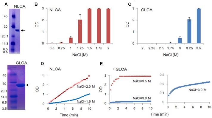
Timely disassembly of viral core composed of self-assembled capsid (CA) in infected host cells is crucial for retroviral replication. Extensive in vitro studies to date on the self-assembly/disassembly mechanism of human immunodeficiency virus type 1 (HIV-1) CA have revealed its core structure and amino acid residues essential for CA-CA intermolecular interaction. However, little is known about in vitro properties of HIV-2 CA. In this study, we comparatively analyzed the polymerization properties of bacterially expressed HIV-1 and HIV-2 CA proteins. Interestingly, a much higher concentration of NaCl was required for HIV-2 CA to self-assemble than that for HIV-1 CA, but once the polymerization started, the reaction proceeded more rapidly than that observed for HIV-1 CA. Analysis of a chimeric protein revealed that N-terminal domain (NTD) is responsible for this unique property of HIV-2 CA. To further study the molecular basis for different in vitro properties of HIV-1 and HIV-2 CA proteins, we determined thermal stabilities of HIV-1 and HIV-2 CA NTD proteins at several NaCl concentrations by fluorescent-based thermal shift assays. Experimental data obtained showed that HIV-2 CA NTD was structurally more stable than HIV-1 CA NTD. Taken together, our results imply that distinct in vitro polymerization abilities of the two CA proteins are related to their structural instability/stability, which is one of the decisive factors for viral replication potential. In addition, our assay system described here may be potentially useful for searching for anti-CA antivirals against HIV-1 and HIV-2.

Keywords: CA-polymerization; CA-stability; Gag-CA; HIV-1; HIV-2; NTD.


Figures

FIGURE 1
FIGURE 1
Structural comparison of NL4-3 and GL-AN Gag-CA proteins. (A) Alignment of NL4-3 and GL-AN Gag-CA sequences. CA amino acid sequences of HIV-1 NL4-3 (GenBank accession number: AF324493) and HIV-2 GL-AN (GenBank accession number: M30895) are aligned. The N-terminal domain (NTD), linker domain, C-terminal domain (CTD), β-hairpin, and helices 1 to 11 (H1 to H11) are shown based on previous studies (Gamble et al., 1996; von Schwedler et al., 2003; Robinson et al., 2014; Gres et al., 2015). (B) Superimposition of the NTD structures. Superposed structures of HIV-1 NTD (green, PDB code: 3H4E) and HIV-2 NTD (gray, PDB code: 2WLV) were depicted by PyMOL ver 1.8.
FIGURE 2
FIGURE 2
In vitro polymerization features of NL4-3 and GL-AN CA proteins. Polymerization reaction was carried out and monitored by OD at 350 nm as described in MATERIALS AND METHODS. (A) SDS-PAGE profile. The purity of CA proteins was checked by SDS-PAGE gel stained with Coomassie Brilliant Blue. Size markers in kDa (on the left) and the CA proteins (arrows) are indicted. NLCA, NL4-3 CA; GLCA, GL-AN CA. (BE) Self-polymerization of CA proteins. Purified CA proteins were added to the buffer containing various concentrations of NaCl, and incubated for 4 h (B–C) or 10 min (D–E) at room temperature. Polymerized products and polymerization kinetics at various concentrations of NaCl were determined by OD at 350 nm for NLCA (B,D) and GLCA (C,E) as shown.
FIGURE 3
FIGURE 3
Tubular structures of in vitro assembled NL4-3 and GL-AN CA proteins. CA proteins were fully polymerized in vitro, fixed by glutaraldehyde, and visualized via TEM as previously described (Sakaguchi et al., 2002; Piroozmand et al., 2006). (A,B) represent micrographs of HIV-1 NL4-3 CA (NLCA) and HIV-2 GL-AN CA (GLCA), respectively. Scale bars: 1 μm in upper panels; 200 nm in lower panels.
FIGURE 4
FIGURE 4
Comparative analysis of several CA proteins for their in vitro polymerization properties. CA-polymerization was performed in vitro and monitored by OD at 350 nm as described in MATERIALS AND METHODS. (A) Polymerization of NL4-3 CA (NLCA), GL-AN CA (GLCA), and NL/GL CA (NL/GL) proteins for 4 h at various NaCl concentrations. The chimeric clone NL/GL has the sequence encoding the NTD of NL4-3 CA and the linker domain/CTD of GL-AN CA (Figure 1A). (B) Polymerization kinetics of NL and NL/GL CA proteins (1.5 M NaCl). (C) Polymerization of NL and GL32NL CA proteins for 4 h at various NaCl concentrations. GL32NL is a chimeric NLCA-derivative clone which has the sequence encoding the very N-terminal region of GL-AN CA (Pro1-Phe32 in Figure 1A).
FIGURE 5
FIGURE 5
Comparison of in vitro polymerization activity of GL-AN and mutant CA proteins. (A) CA–CA interacting surface of HIV-1. Two CA molecules (greenish and bluish) are shown. Three amino acids (M39, T54, and T58 in the H2–H3 region) critical for the interaction are highlighted as shown (see Pornillos et al., 2009, for details). The structure of HIV-1 CA (PDB code: 3H4E) was depicted by PyMOL ver 1.8. (B) Polymerization of parental and mutant CA proteins. CA-polymerization was performed in vitro for 4 h at various NaCl concentrations, and monitored by OD at 350 nm as described in the Section “Materials and Methods”. The mutant clone GLmtCA has three amino acid substitutions relative to GLCA as indicated.
FIGURE 6
FIGURE 6
Thermal stability of CA NTD proteins derived from HIV-1 NL4-3, and HIV-2 GL-AN. The thermal stability of NLCA and GLCA NTD proteins in the presence of 250 mM NaCl was determined by DSF as described in the Section “Materials and Methods”. SYPRO orange fluorescence intensity (FI) at varying temperatures (upper panel) and derivative melt curves calculated by differences in FI at each temperature (lower panel) are shown. Peak temperatures in the curves (dFI/dT) were considered as Tm.
FIGURE 7
FIGURE 7
Thermal stability of CA NTD proteins at various NaCl concentrations. The thermal stability of NLCA and GLCA NTD proteins at various NaCl concentrations was evaluated as described in the legend to Figure 6. FI values and melt curves (A,B, respectively), and Tm shifts induced by NaCl (C) are shown.

Similar articles

Cited by

References

    1. Adachi A., Gendelman H. E., Koenig S., Folks T., Willey R., Rabson A., et al. (1986). Production of acquired immunodeficiency syndrome-associated retrovirus in human and nonhuman cells transfected with an infectious molecular clone. J. Virol. 59 284–291. - PMC - PubMed
    1. Alfadhli A., Dhenub T. C., Still A., Barklis E. (2005). Analysis of human immunodeficiency virus type 1 Gag dimerization-induced assembly. J. Virol. 79 14498–14506. 10.1128/JVI.79.23.14498-14506.2005 - DOI - PMC - PubMed
    1. Barklis E., Alfadhli A., McQuaw C., Yalamuri S., Still A., Barklis R. L., et al. (2009). Characterization of the in vitro HIV-1 capsid assembly pathway. J. Mol. Biol. 387 376–389. 10.1016/j.jmb.2009.01.058 - DOI - PMC - PubMed
    1. Bhattacharya A., Alam S. L., Fricke T., Zadrozny K., Sedzicki J., Taylor A. B., et al. (2014). Structural basis of HIV-1 capsid recognition by PF74 and CPSF6. Proc. Natl. Acad. Sci. U.S.A. 111 18625–18630. 10.1073/pnas.1419945112 - DOI - PMC - PubMed
    1. Black L. R., Aiken C. (2010). TRIM5alpha disrupts the structure of assembled HIV-1 capsid complexes in vitro. J. Virol. 84 6564–6569. 10.1128/JVI.00210-10 - DOI - PMC - PubMed

LinkOut - more resources