. 2017 Jun 14:8:1085.
doi: 10.3389/fmicb.2017.01085. eCollection 2017.

A Novel Method to Titrate Herpes Simplex Virus-1 (HSV-1) Using Laser-Based Scanning of Near-Infrared Fluorophores Conjugated Antibodies

Affiliations

A Novel Method to Titrate Herpes Simplex Virus-1 (HSV-1) Using Laser-Based Scanning of Near-Infrared Fluorophores Conjugated Antibodies

Marco Fabiani et al. Front Microbiol. .

Abstract

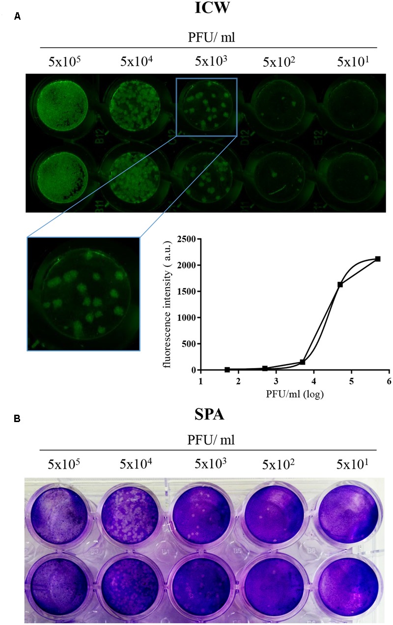
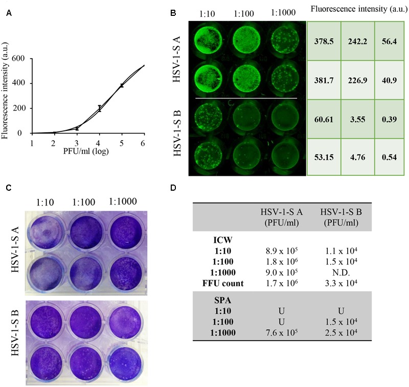
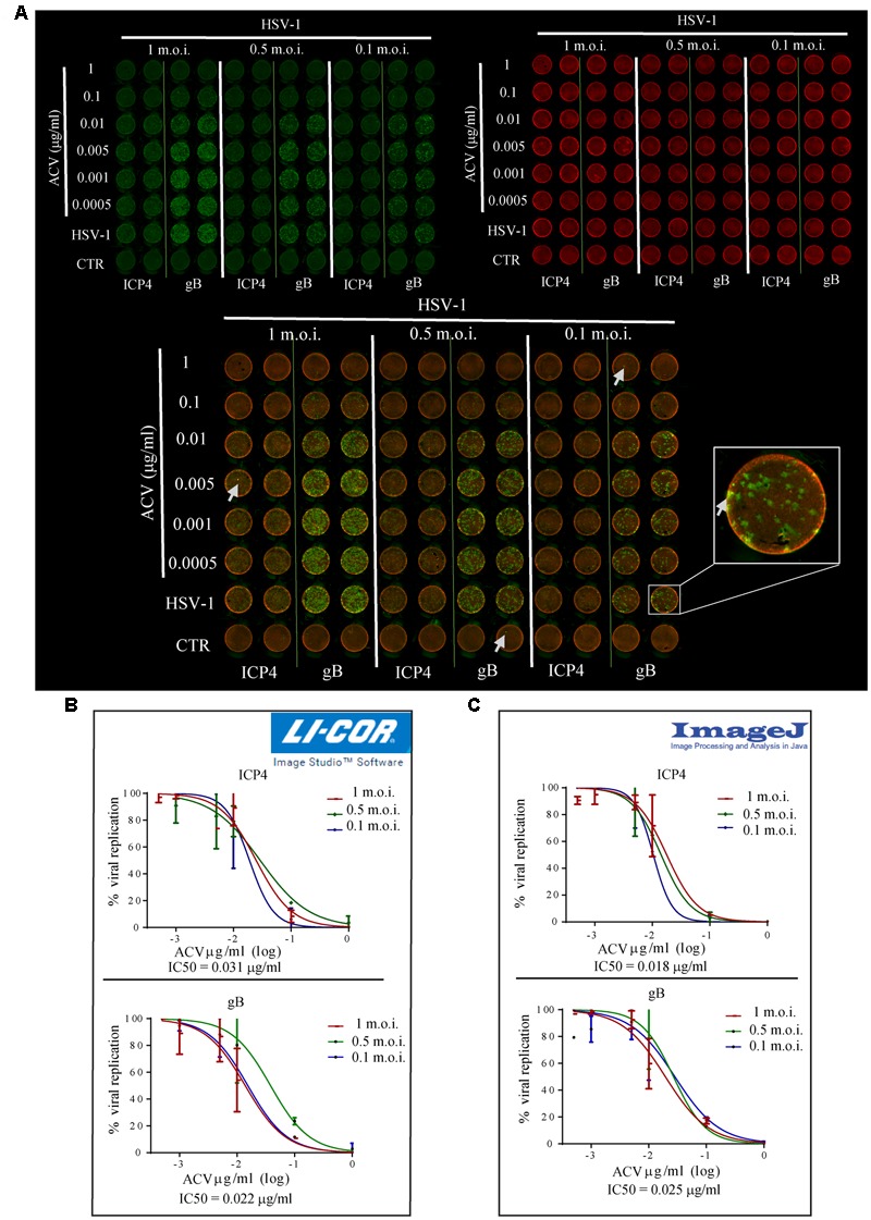
Among several strategies used for Herpes simplex virus (HSV) detection in biological specimens, standard plaque assay (SPA) remains the most reliable method to evaluate virus infectivity and quantify viral replication. However, it is a manual procedure, thereby affected by operator subjectivity, and it may be particularly laborious for multiple sample analysis. Here we describe an innovative method to perform the titration of HSV type 1 (HSV-1) in different samples, using the "In-Cell WesternTM" Assay (ICW) from LI-COR, a quantitative immunofluorescence assay that exploits laser-based scanning of near infrared (NIR). In particular, we employed NIR-immunodetection of viral proteins to monitor foci of HSV-1 infection in cell monolayers, and exploited an automated detection of their fluorescence intensity to evaluate virus titre. This innovative method produced similar and superimposable values compared to SPA, but it is faster and can be performed in 96 well plate, thus allowing to easily and quickly analyze and quantify many samples in parallel. These features make our method particularly suitable for the screening and characterization of antiviral compounds, as we demonstrated by testing acyclovir (ACV), the main anti-HSV-1 drug. Moreover, we developed a new data analysis system that allowed to overcome potential bias due to unspecific florescence signals, thus improving data reproducibility. Overall, our method may represents a useful tool for both clinical and research purposes.

Keywords: HSV-1; antivirals; herpes simplex virus; immunostaining; near-infrared fluorescence; plaque assay; virus titration.


Figures

FIGURE 1
FIGURE 1
“In-Cell WesternTM” Assay is a suitable technique to detect HSV-1 infection in cells. (A) Vero cells were seeded in 96 wells and infected with the indicated 10-fold serial dilutions of HSV-1. 24 h p.i. cells were fixed and immunostained with anti-gB antibody, followed by incubation with IRDye 800CW goat anti-mouse antibody, and relative fluorescence detected with LI-COR Odyssey Infrared Imaging System. A representative image of one of the three experiments performed is shown, with a close up image of HSV-1 foci of infection (inset) and the relative standard curve built up with the mean value of fluorescence intensity (arbitrary unit, a.u.) detected for each experimental point. (B) Representative image of SPA performed with the same HSV-1 dilutions used in (A).
FIGURE 2
FIGURE 2
“In-Cell WesternTM” Assay vs. standard plaque assay. (A) ICW Internal standard curve built up by using the mean value of intensity fluorescence detected in Vero cells infected with 10-fold serial dilutions of HSV-1 with a known titre. (B,C) Two unknown HSV-1 samples (HSV-1-SA and HSV-1-SB) were titrated using ICW (B) and SPA (C) as described in Section “Materials and Methods.” (D) Mean values of HSV-1-SA and HSV-1-SB titer for each tested dilution in the two assays. N.D. = not detectable; U = uncountable.
FIGURE 3
FIGURE 3
“In-Cell WesternTM” Assay is a suitable technique for antiviral screening. (A) HSV-1 infected Vero cells (with the indicated m.o.i.) were treated for 24 h with concentrations of ACV ranging from 0.0005 to 1 μg/ml. Fixed cells were incubated with anti-gB or anti-ICP4 antibodies and then stained with IRDye 800 CW (green fluorescence) and CellTag 700 Stain (red fluorescence). Arrows highlight unspecific fluorescence peaks (a close up view in the inset). (B,C) Representative dose-response curves of ACV activity on HSV-1 replication (shown as percentage of virus replication in ACV treated cells vs. untreated ones) using LI-COR Image Studio Software (B) or Image-J (C) for data analysis. ACV IC50 was calculated from each staining and the mean values resulted from experiments performed with different m.o.i. of virus are reported (0.031 μg/ml for ICP4 staining, 0.022 μg/ml for gB staining using LI-COR Image Studio Software; 0.018 μg/ml for ICP4 staining, 0.025 μg/ml for gB staining using Image-J software).

Similar articles

Cited by

References

    1. Baer A., Kehn-Hall K. (2014). Viral concentration determination through plaque assays: using traditional and novel overlay systems. J. Vis. Exp. 93:52065 10.3791/52065 - DOI - PMC - PubMed
    1. Baker D. A., Gonik B., Milch P. O., Berkowitz A., Lipson S., Verma U. (1989). Clinical evaluation of a new herpes simplex virus ELISA: a rapid diagnostic test for herpes simplex virus. Obstet. Gynecol. 73(3 Pt 1) 322–325. 10.1016/0020-7292(89)90375-5 - DOI - PubMed
    1. Borisevich V., Nistler R., Hudman D., Yamshchikov G., Seregin A., Yamshchikov V. (2007). A highly sensitive and versatile virus titration assay in the 96-well microplate forma. J. Virol. Methods 147 197–205. 10.1016/j.jviromet.2007.08.015 - DOI - PubMed
    1. Burnett L. C., Lunn G., Coico R. (2009). Biosafety: guidelines for working with pathogenic and infectious microorganisms. Curr. Protoc. Microbiol. 69 A.1V.1–A.1V.8. 10.1002/9780471729259.mc01a01s13 - DOI - PMC - PubMed
    1. Civitelli L., Panella S., Marcocci M. E., De Petris A., Garzoli S., Pepi F., et al. (2014). In vitro inhibition of herpes simplex virus type 1 replication by Mentha suaveolens essential oil and its main component piperitenone oxide. Phytomedicine 21 857–865. 10.1016/j.phymed.2014.01.013 - DOI - PubMed

LinkOut - more resources