. 2016 Apr 21;7(4):2470-2491.
doi: 10.1039/c5sc03224e. Epub 2015 Dec 23.

The rise of 3-d single-ion magnets in molecular magnetism: towards materials from molecules?

Affiliations

The rise of 3-d single-ion magnets in molecular magnetism: towards materials from molecules?

Jamie M Frost et al. Chem Sci. .

Abstract

Single-molecule magnets (SMMs) that contain one spin centre (so-called single-ion magnets) theoretically represent the smallest possible unit for spin-based electronic devices. The realisation of this and related technologies, depends on first being able to design systems with sufficiently large energy barriers to magnetisation reversal, Ueff, and secondly, on being able to organise these molecules into addressable arrays. In recent years, significant progress has been made towards the former goal - principally as a result of efforts which have been directed towards studying complexes based on highly anisotropic lanthanide ions, such as Tb(iii) and Dy(iii). Since 2013 however, and the remarkable report by Long and co-workers of a linear Fe(i) system exhibiting Ueff = 325 K, single-ion systems of transition metals have undergone something of a renaissance in the literature. Not only do they have important lessons to teach us about anisotropy and relaxation dynamics in the quest to enhance Ueff, the ability to create strongly coupled spin systems potentially offers access to a whole of host of 1, 2 and 3-dimensional materials with interesting structural and physical properties. This perspective summarises recent progress in this rapidly expanding sub-genre of molecular magnetism from the viewpoint of the synthetic chemist, with a particular focus on the lessons that have so far been learned from single-ion magnets of the d-block, and, the future research directions which we feel are likely to emerge in the coming years.


Figures

Fig. 1
Fig. 1. Double-well energy diagram for negative (left) and positive (right) D.
Fig. 2
Fig. 2. Schematic representation of possible relaxation pathways in SMMs. Blue lines represent spin states. The grey line represents a virtual state by which Raman relaxation proceeds. Colour code: green = ground state QTM, red = thermally assisted QTM (TA-QTM), purple = Orbach relaxation, grey = Raman relaxation.
Fig. 3
Fig. 3. (a) Molecular structure of Hpy[Fe17O16(OH)12(py)12Cl4]Cl4 (b) metallic core of Hpy[Fe17O16(OH)12(py)12Cl4]Cl4 and (c) a portion of the structure of magnetite demonstrating its similarities with Hpy[Fe17O16(OH)12(py)12Cl4]Cl4. Colour code: orange (Fe), red (O), blue (N), green (Cl), grey (C). Hydrogen atoms omitted for clarity. Adapted from ref. 19.
Fig. 4
Fig. 4. Molecular structure of some multi-decker 4f COT complexes. Colour code: yellow (Ln), purple (K), green (Si), red (O), grey (C). Hydrogen atoms omitted for clarity.
Fig. 5
Fig. 5. A hypothesised chain-like arrangement of M(COT)2 monomers with uniaxial anisotropy, illustrating the concept of modular design of single-chain magnets. The axial anisotropy of each monomer is depicted as blue vectors and the vector addition of the monomeric axial anisotropies yields the net axial anisotropy (purple).
Fig. 6
Fig. 6. Energy level diagram illustrating the effect of a weak octahedral crystal field and spin orbit coupling on the Ni(ii) ion as described in the text. Levels expanded for clarity. The multiplicity of states arising from spin–orbit coupling are given in brackets.
Fig. 7
Fig. 7. Energy level diagram illustrating the effect of a weak octahedral crystal field and spin orbit coupling on the Co(ii) ion as described in the text. Levels expanded for clarity. The multiplicity of states arising from spin–orbit coupling are given in brackets.
Fig. 8
Fig. 8. (a) Molecular structure of [Fe(1-ptz)6][(BF4)]2 (2). Colour code: orange (Fe), blue (N), and grey (C). Hydrogen atoms omitted for clarity. (b) Excitation and de-excitation cycling between the HS and LS configuration of 2, represented as a variation in the χMT product at 10 K, under a 5000 Oe field. Reprinted with permission from ref. 28. Copyright (2013) American Chemical Society.
Fig. 9
Fig. 9. Molecular structures of four coordinate trigonal pyramidal Fe(ii) complexes of the form [(tpaR)Fe]. Colour code: orange (Fe), green (Cl), blue (N), grey (C). Hydrogen atoms omitted for clarity.
Fig. 10
Fig. 10. (a) Molecular structure of [Fe(N(TMS)2)2(PCy3)], (TMS = SiMe3, Cy = cyclohexyl) (8). Colour code: orange (Fe), plum (P), teal (Si), blue (N), grey (C). Hydrogen atoms omitted for clarity. (b) TD-DFT calculated excited states and β-spin molecular orbitals (MOs). The energies of the excited states and the metal contribution to the MOs are also shown. Reprinted with permission from ref. 34. Copyright (2011) American Chemical Society.
Fig. 11
Fig. 11. Molecular structures of linear two coordinate Fe(ii) complexes (from left to right): Fe[N(SiMe3)(Dipp)]2 (11), Fe[C(SiMe3)3]2 (12), Fe[N(H)Ar′]2 (13), Fe[N(H)Ar*]2 (14), Fe[OAr′]2 (15), and Fe[N(H)Ar#]2 (16). Colour code: orange (Fe), teal (Si), blue (N), red (O), grey (C). Hydrogen atoms omitted for clarity. Below is the frequency dependence of the out-of-phase magnetic susceptibility (χ′′) as a function of temperature for each complex. Data were collected under applied dc fields of 500 Oe (11), 500 Oe (12), 875 Oe (13), 875 Oe (14), 2500 Oe (15), and 1000 Oe (16), respectively. Reprinted with permission from ref. 36b. Copyright (2013) Royal Society of Chemistry.
Fig. 12
Fig. 12. Molecular structure of [Co(SCN)2(4-dzbpy)4] (18) (left) and out-of-phase magnetic susceptibility as a function of temperature (right), with an inset showing the Arrhenius fit of the data. Reprinted with permission from ref. 39. Copyright (2003) American Chemical Society.
Fig. 13
Fig. 13. (a) Molecular structure of [HNEt3][CoIICoIII3L6] (19). Colour code: dark purple (Co(ii)), light purple (Co(iii)), red (O), blue (N), grey (C) and light green (Br). Hydrogen atoms omitted for clarity. (b) Simplified d-orbital splitting diagram (c) simplified coordination environment of central Co(ii) ion and (d) Out-of-phase magnetic susceptibility for 19 under zero applied dc field. Arrhenius plot of natural log of the relaxation time vs. inverse temperature as inset. Reprinted with permission from ref. 42. Copyright (2013) Royal Society of Chemistry.
Fig. 14
Fig. 14. Out-of-phase magnetic susceptibility for [Co(Pzox)3(BC6H5)]Cl (20) measured under; (a) zero applied dc field and (b) an applied dc field of 1500 Oe. (c) Arrhenius plot of the natural log of the relaxation time vs. inverse temperature under a zero applied dc field and (d) an applied dc field of 1500 Oe. Reprinted with permission from ref. 43. Copyright (2015) American Chemical Society.
Fig. 15
Fig. 15. Molecular structures of [CoIIICoII(LH2)2(X)(H2O)](H2O)4 and the frequency dependence of the out-of-phase magnetic susceptibility collected in a 1000 Oe dc field. (a) X = Cl (21) and (b) X = Br (22). Colour code: purple (Co), bright green (Cl), light green (Br), blue (N), red (O), grey (C). Hydrogen atoms omitted for clarity. Reprinted with permission from ref. 44. Copyright (2014) American Chemical Society.
Fig. 16
Fig. 16. (a) Molecular structures of [Co(LPh)(NCS)2] (25, left) and [Co(LMe)(NCS)2] (24, right). Colour code: purple (Co), blue (N), yellow (S), grey (C). (b) d-orbital splitting diagrams for 24 and 25, highlighting the effect of metal ion displacement from the basal plane. Reprinted with permission from ref. 47. Copyright (2011) American Chemical Society.
Fig. 17
Fig. 17. Energy level diagram depicting selected β-spin frontier molecular orbitals of [Co(terpy)Cl2] (Cs symmetry) (26), [Co(terpy)(NCS)2] (C2v symmetry) (27) and [Co(terpy)2]2+ (Cs symmetry). The increase in the number of β-spins for [Co(terpy)2]2+ comes at the cost of an α-spin, resulting in an overall decrease in the molecular spin state. Reprinted with permission from ref. 48. Copyright (2013) Wiley-VCH.
Fig. 18
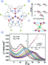
Fig. 18. (a) Molecular structure, d-orbital splitting diagram and χ′′ vs. ν plot as a function of H for [Co(SePh)4]2– (32). Colour code: purple (Co), yellow (S), grey (C). Counter cation and hydrogen atoms omitted for clarity. Reprinted with permission from ref. 50. Copyright (2011) American Chemical Society. (b) Molecular structure and absorption spectra changes under UV irradiation, thus demonstrating the photochromic behaviour of [Co(hpbdti)2] (hpbdtiH = 2-(2-hydroxpheyl)-4,5-bis(2,5-dimethyl(3-thienyl))-1H-imidazole) (37). Colour code: purple (Co), yellow (S), red (O), blue (N), grey (C). Hydrogen atoms omitted for clarity. Reprinted with permission from ref. 54a. Copyright (2013) Royal Society of Chemistry.
Fig. 19
Fig. 19. Molecular structure and d-orbital splitting diagram for the three coordinate Co(ii) complexes, [Li(15-crown-5)][Co{N(SiMe3)2}3] (46), [Co{N(SiMe3)2}2(THF)] (47) and [Co{N-(SiMe3)2}2(PCy3)] (48). Colour code: purple (Co), plum (P), teal (Si), blue (N), red (O), grey (C). Hydrogen atoms omitted for clarity. Adapted with permission from ref. 56. Copyright (2014) American Chemical Society.
Fig. 20
Fig. 20. (a) Molecular structure of [MnIII(5-TMAM(R)-salmen)(H2O)CoIII(CN)6]·7H2O·MeCN (49) Colour code: teal (Mn), purple (Co), blue (N), red (O), bright green (Cl), grey (C). Hydrogen atoms omitted for clarity. To the right of this, is the temperature dependence of the ac susceptibilities (χ′ and χ′′) under an applied dc field of 4500 Oe and an ac field of 5 Oe. Reprinted with permission from ref. 57. Copyright (2013) American Chemical Society. (b) Molecular structure of Ph4P[Mn(opbaCl2)(py)2] (50). Colour code: teal (Mn), blue (N), red (O), bright green (Cl), grey (C). Hydrogen atoms omitted for clarity. To the right of this, is the temperature dependence of the out-of-phase magnetic susceptibility (χ′′) under an applied dc field of 1000 Oe and 4 Oe oscillating field (inset: Arrhenius plot), and, the tweep rate dependence of the magnetisation at 0.5 K (inset: at 0.03 K). Reprinted with permission from ref. 58. Copyright (2013) Wiley-VCH.
Fig. 21
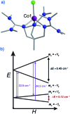
Fig. 21. (a) Molecular structure of [(3G)CoCl] (CF3SO3) (58). Colour code: purple (Co), bright green (Cl), blue (N), grey (C). Hydrogen atoms omitted for clarity. (b) Zeeman splitting diagram for 58, where the red arrows indicated the direct relaxation process, the purple arrows correspond to the excitation energies related to the Orbach processes, and the blue arrows correspond to the relaxation via the Orbach process. All values presented were determined for S = 3/2, g, D, and E as determined by EPR spectroscopy. Reprinted with permission from ref. 65. Copyright (2012) Royal Society of Chemistry.
Fig. 22
Fig. 22. Molecular structure of [Co(L)(OAc)Y(NO3)2] (65), with fitting of the relaxation time to a Tn power law as described in the text. Colour code: purple (Co), yellow (Y), blue (N), red (O), grey (C). Hydrogen atoms omitted for clarity. Reprinted with permission from ref. 71. Copyright (2013) Wiley-VCH.
Fig. 23
Fig. 23. Frequency dependent out-of-phase magnetic susceptibility (χ′′) as a function of applied field (200–8200 Oe) at 2 K of [Co(DAPBH)(NO3)(H2O)] (67) magnetically diluted with [Zn(DAPBH)(NO3)(H2O)]. A and B represent two relaxation processes. Colour code: purple (Co) and yellow (Zn). Reprinted with permission from ref. 73. Copyright (2015) Royal Society of Chemistry.
None
Jamie M. Frost
None
Katie L. M. Harriman
None
Muralee Murugesu

References

    1. Buschowand K. H. J. and de Boer F. R., Physics of Magnetic Materials, World Scientific, 1985.
    1. Craig G. A., Murrie M. Chem. Soc. Rev. 2015;44:2135. - PubMed
    2. Gómez-Coca S., Aravena D., Morales R., Ruiz E. Coord. Chem. Rev. 2015;290:379–392.
    1. Bogani L., Wernsdorfer W. Nat. Mater. 2008;7:179. - PubMed
    1. Sessoli R., Gatteschi D., Caneschi A., Novak M. A. Nature. 1993;365:141.
    1. Furrer A., Waldmann O. Rev. Mod. Phys. 2013;85:367.
    2. Timco G. A., McInnes E. J. L., Winpenny R. E. P. Chem. Soc. Rev. 2013;42:1796. - PubMed
    3. Timco G. A., Faust T. B., Tuna F., Winpenny R. E. P. Chem. Soc. Rev. 2011;40:3067. - PubMed
    4. Santini P., Carretta S., Troiani F., Amoretti G. Phys. Rev. Lett. 2011;107:230502. - PubMed

LinkOut - more resources