. 2016 Apr 21;7(4):2553-2562.
doi: 10.1039/c5sc04197j. Epub 2016 Jan 20.

A new Pseudomonas quinolone signal (PQS) binding partner: MexG

Affiliations

A new Pseudomonas quinolone signal (PQS) binding partner: MexG

James T Hodgkinson et al. Chem Sci. .

Abstract

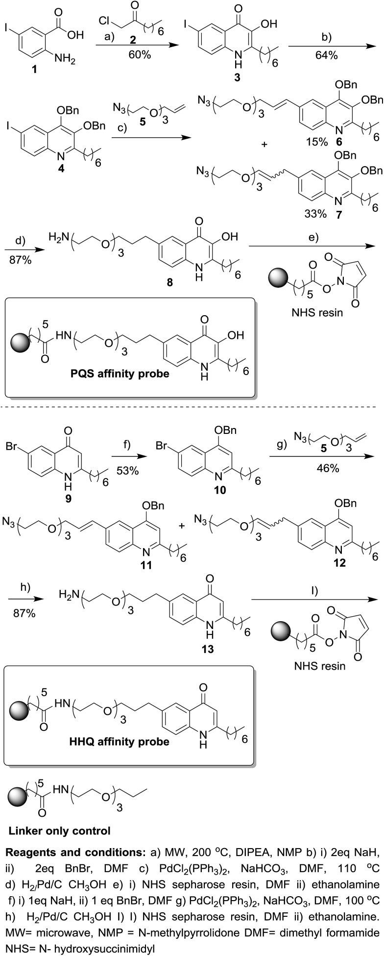
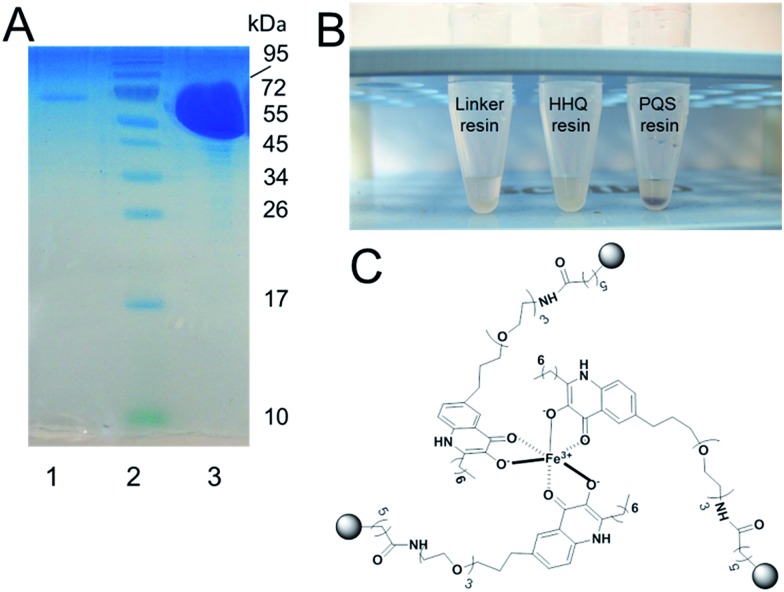
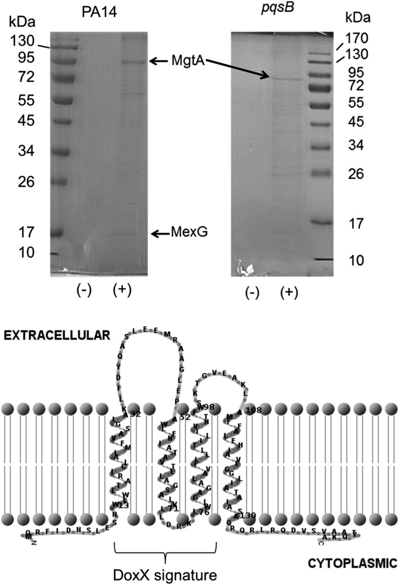
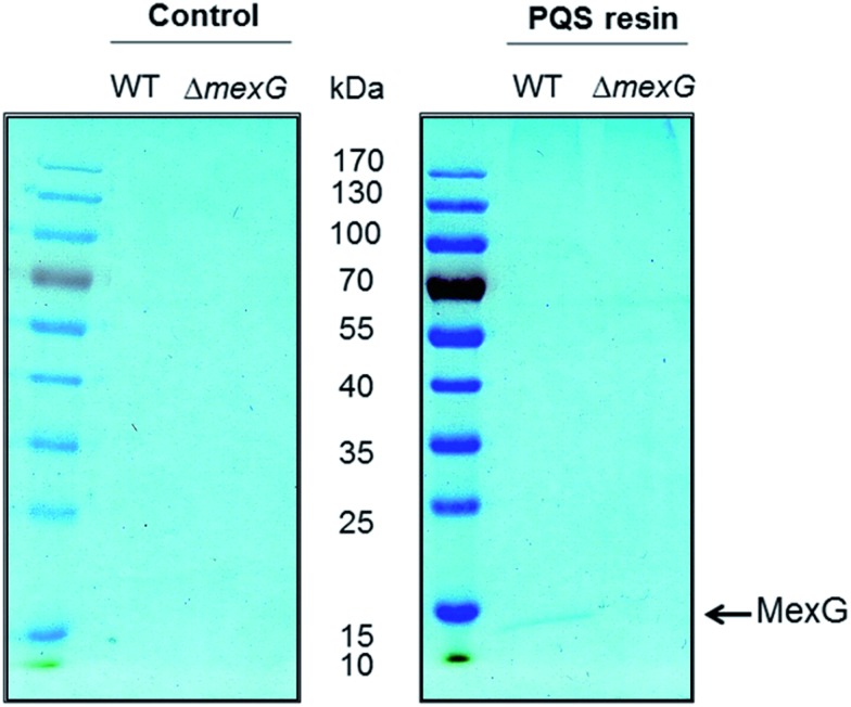
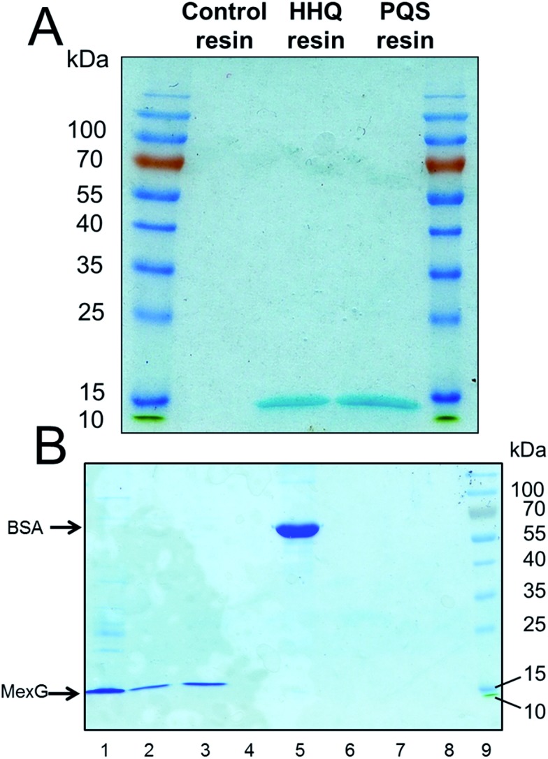
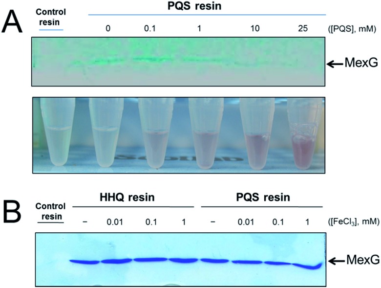
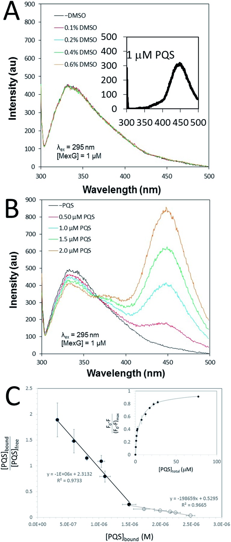
The opportunistic pathogen Pseudomonas aeruginosa utilises the cell-cell signalling mechanism known as quorum sensing to regulate virulence. P. aeruginosa produces two quinolone-based quorum sensing signalling molecules; the Pseudomonas quinolone signal (PQS) and its biosynthetic precursor 2-heptyl-4(1H)-quinolone (HHQ). To date, only one receptor (the PqsR protein) has been identified that is capable of binding PQS and HHQ. Here, we report on the synthesis of PQS and HHQ affinity probes for chemical proteomic studies. The PQS affinity probe very effectively captured PqsR in vitro. In addition, we also identified an interaction between PQS and the "orphan" RND efflux pump protein, MexG. The PQS-MexG interaction was further confirmed by purifying MexG and characterizing its ability to bind PQS and HHQ in vitro. Our findings suggest that PQS may have multiple binding partners in the cell and provide important new tools for studying quinolone signalling in P. aeruginosa and other organisms.


Figures

Scheme 1
Scheme 1. Synthesis of HHQ and PQS affinity probes. For full experimental details on the preparation of intermediates and the linker only control see ESI.
Fig. 1
Fig. 1. The PQS affinity probe retains affinity for PqsRLBD and Fe3+. (A) Purified PqsR ligand binding domain (PqsRLBD) binds strongly to immobilized PQS but not to the control linker resin. Purified MBP-tagged PqsRLBD was incubated with aliquots of the control or PQS-derivatized resin, see ESI. The resin samples were extensively washed and the bound protein was eluted by boiling the washed beads in SDS sample buffer. The panel shows a Coomassie Brilliant Blue-stained SDS-PAGE gel of the protein eluting from control beads (lane 1) or PQS-derivatized beads (lane 3). Molecular mass markers are shown in lane 2. (B) Aliquots (50 μL) of control beads, HHQ beads or PQS beads (as indicated) were mixed with 1 mM FeCl3. The PQS beads immediately turned deep red due to the formation of the PQS–iron complex. (C) Proposed structure of the PQS–Fe complex on probe resulting in dark red colouration.
Fig. 2
Fig. 2. The PQS affinity probe captures MgtA and MexG from the detergent-solubilized cell membrane fraction. Detergent-solubilized cell membranes from stationary phase cultures of wild-type PA14 or an isogenic pqsB mutant, as indicated, were mixed with aliquots of either control resin (–) or PQS resin (+). Following extensive washing of the resins, bound proteins were eluted by boiling the resin samples in SDS sample buffer. The eluted proteins were resolved by SDS-PAGE and an image of the Coomassie Brilliant Blue-stained gel is shown. Following staining of the gel, a selection of bands were excised and identified by MALDI-MS. The MgtA and MexG-containing bands are indicated. Predicted membrane topology of MexG. The DoxX domain (residues 15–95, encompassing most of helices 1–3) signature is indicated. The MexG amino acid sequence was analysed by TMHMM and the output was depicted using TMRPRES2D.
Fig. 3
Fig. 3. Deletion of the DoxX motif abolishes MexG binding to PQS. Detergent-solubilized membranes from either wild-type PAO1 or from an in-frame mexG deletion mutant (as indicated) were mixed with control resin or PQS-derivatized resin and incubated to allow binding. The resins were washed extensively and the bound proteins were eluted by boiling in SDS sample buffer. The figure shows a Coomassie Brilliant Blue-stained gel of the proteins bound to control resin (left panel) and PQS resin (right panel). Note that essentially the only protein detectable in this experiment was MexG from the wild-type extract bound to the PQS resin. Control = linker only resin.
Fig. 4
Fig. 4. Purified MexG binds to immobilized PQS and immobilized HHQ in vitro. (A) Purified MexG was incubated with either linker resin, HHQ or PQS affinity probes (as indicated) for 1 h. After extensive washing of the resins, the bound protein was eluted in SDS sample buffer and resolved by SDS-PAGE. The resulting Commassie Brilliant Blue-stained gel is shown. (B) The experiment in (A) was repeated using BSA in place of MexG. Lane 1; an aliquot of the MexG sample that was loaded onto the resin, lane 2; MexG bound to PQS resin, lane 3; MexG bound to HHQ resin, lane 4; MexG bound to control resin, lane 5; BSA alone, lane 6; BSA bound to PQS resin, lane 7; BSA bound to HHQ resin, lane 8; BSA bound to control resin, lane 9; molecular mass markers.
Fig. 5
Fig. 5. Soluble PQS outcompetes MexG bound to the PQS affinity probe. (A) Purified MexG was incubated with PQS resin or control resin (as indicated) in the presence of increasing concentrations of “free” PQS. The bound protein remaining after extensive washing of the resins was visualised by SDS-PAGE. The upper panel shows the Commassie Brilliant Blue-stained SDS-PAGE gel. The lower panel shows Eppendorf tubes containing the unbound material after incubating with the resin (but before the wash steps) mixed with FeCl3. Note the increased intensity of red Fe3+–PQS complex formed at higher “free” PQS concentrations correlates with a reduction in the amount of MexG bound to the resin after washing. (B) Purified MexG was inclubated with control resin, HHQ resin or PQS resin (as indicated) in the presence of the indicated concentrations of FeCl3. Bound proteins were resolved by SDS-PAGE and the image shows the resulting Coomassie Brilliant Blue-stained gel.
Fig. 6
Fig. 6. Fluorescence emission spectra of MexG and PQS. (A) The fluorescence emission spectra of purified MexG (1 μM final concentration) was measured in the presence of the indicated amounts of added DMSO (DMSO was used as a solvent for PQS). The excitation wavelength was 295 nm. The highest final concentration of DMSO tested (0.6% v/v) was greater than the highest DMSO concentration achieved during the titration of PQS. Note that the addition of DMSO does not change the emission spectrum of MexG. Inset. Emission spectrum of 1 μM PQS excited at 295 nm. (B) Emission spectrum of purified MexG in the presence of increasing concentrations of PQS. The excitation wavelength was 295 nm. Note how increasing PQS concentrations lead to an increase in emission at ca. 450 nm and a concomitant decrease (quenching) of emission at ca. 340 nm. (C) Quantitation of PQS binding to MexG. Scatchard plot of the binding data derived from fluorescence quenching experiments. Note the biphasic binding behaviour of the MexG:PQS interaction, indicative of tight (filled symbols) and weaker (open symbols) binding interactions. The slope of each linear regression yields –1/Kd. Inset. The figure shows the fraction of protein fluorescence that was quenched at different total concentrations of added PQS.

References

    1. Waters C. M., Bassler B. L. Annu. Rev. Cell Dev. Biol. 2005;21:319. - PubMed
    2. Geske G. D., O'Neil J. C., Blackwell H. E. Chem. Soc. Rev. 2008;37:1432. - PMC - PubMed
    3. Whitehead N. A., Barnard A. M., Slater H., Simpson N. J., Salmond G. P. FEMS Microbiol. Rev. 2001;25:365. - PubMed
    1. Galloway W. R. D. J., Hodgkinson J. T., Bowden S. D., Welch M., Spring D. R. Chem. Rev. 2011;111:28. - PubMed
    2. Dickschat J. S. Nat. Prod. Rep. 2010;27:343. - PubMed
    1. Atkinson S., Williams P. J. R. Soc., Interface. 2009;6:959. - PMC - PubMed
    2. Antunes L. C., Ferreria R. B., Buckner M. M., Finlay B. B. Microbiology. 2010;156:2271. - PubMed
    1. Jimenez P. N., Koch G., Thompson J. A., Xavier K. B., Cool R. H., Quax W. J. Microbiol. Mol. Biol. Rev. 2012;76:46. - PMC - PubMed
    2. Laverty G., Gorman S. P., Gilmore B. F. Pathogens. 2014;3:596. - PMC - PubMed
    1. Lintz M. J., Oinuma K., Wysoczynski C. L., Greenberg E. P., Churchill M. E. Proc. Natl. Acad. Sci. U. S. A. 2011;108:15763. - PMC - PubMed

LinkOut - more resources