

Coupling functionalized cobalt ferrite nanoparticle enrichment with online LC/MS/MS for top-down phosphoproteomics
- PMID: 28660060
- PMCID: PMC5472028
- DOI: 10.1039/c6sc05435h
Coupling functionalized cobalt ferrite nanoparticle enrichment with online LC/MS/MS for top-down phosphoproteomics
Abstract
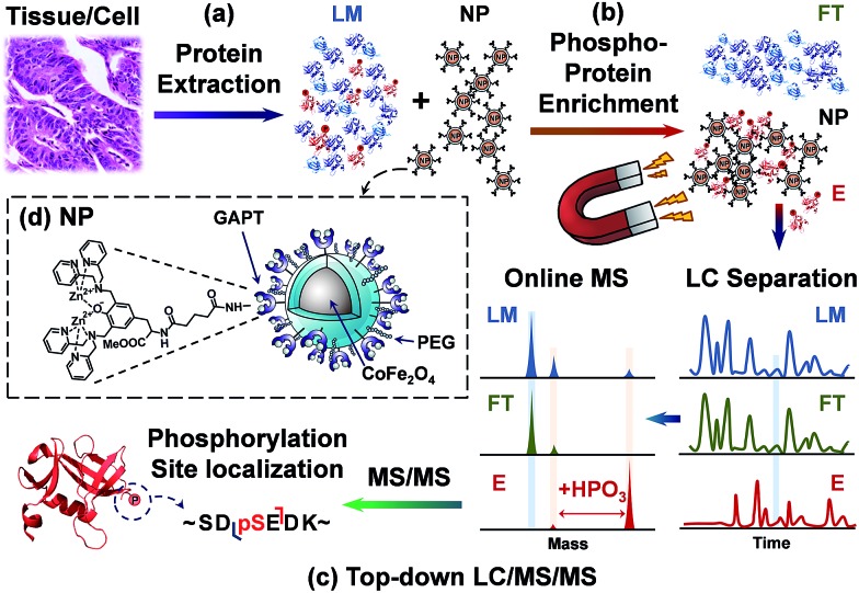
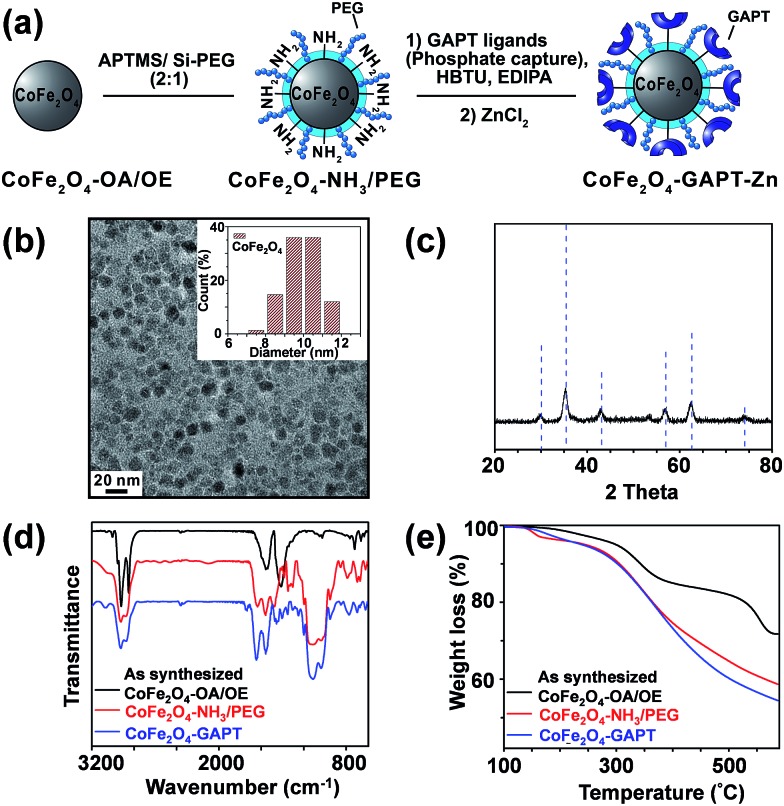
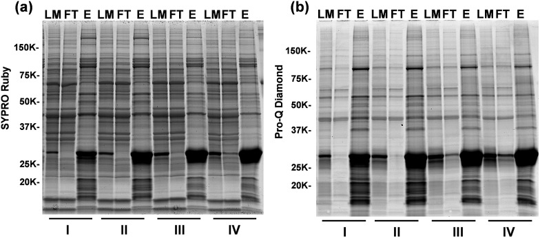
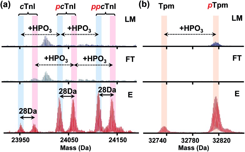
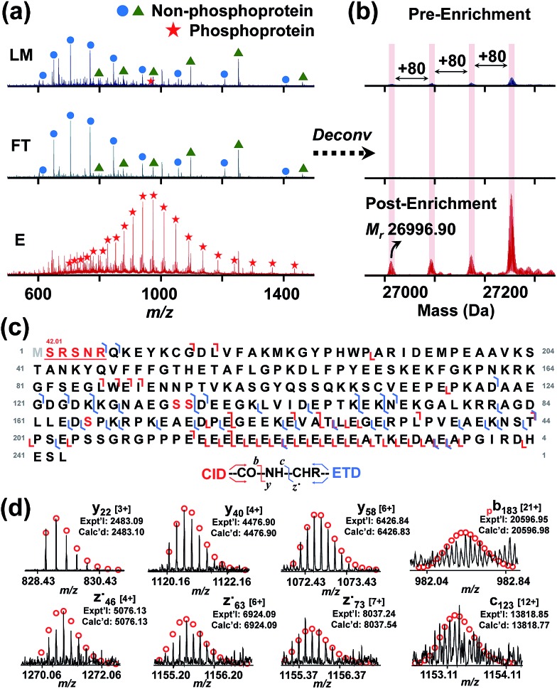
Phosphorylation plays pivotal roles in cellular processes and dysregulated phosphorylation is considered as an underlying mechanism in many human diseases. Top-down mass spectrometry (MS) analyzes intact proteins and provides a comprehensive analysis of protein phosphorylation. However, top-down MS-based phosphoproteomics is challenging due to the difficulty in enriching low abundance intact phosphoproteins as well as separating and detecting the enriched phosphoproteins from complex mixtures. Herein, we have designed and synthesized the next generation functionalized superparamagnetic cobalt ferrite (CoFe2O4) nanoparticles (NPs), and have further developed a top-down phosphoproteomics strategy coupling phosphoprotein enrichment enabled by the functionalized CoFe2O4 NPs with online liquid chromatography (LC)/MS/MS for comprehensive characterization of phosphoproteins. We have demonstrated the highly specific enrichment of a minimal amount of spike-in β-casein from a complex tissue lysate as well as effective separation and quantification of its phosphorylated genetic variants. More importantly, this integrated top-down phosphoproteomics strategy allows for enrichment, identification, quantification, and comprehensive characterization of low abundance endogenous phosphoproteins from complex tissue extracts on a chromatographic time scale.
Figures

Similar articles
-
Reproducible Large-Scale Synthesis of Surface Silanized Nanoparticles as an Enabling Nanoproteomics Platform: Enrichment of the Human Heart Phosphoproteome.Nano Res. 2019 Jun;12(6):1473-1481. doi: 10.1007/s12274-019-2418-4. Epub 2019 May 29. Nano Res. 2019. PMID: 31341559 Free PMC article.
-
Integrated workflow for characterizing intact phosphoproteins from complex mixtures.Anal Chem. 2009 Jun 1;81(11):4210-9. doi: 10.1021/ac802487q. Anal Chem. 2009. PMID: 19425582 Free PMC article.
-
Combining Metabolic ¹⁵N Labeling with Improved Tandem MOAC for Enhanced Probing of the Phosphoproteome.Methods Mol Biol. 2015;1306:81-96. doi: 10.1007/978-1-4939-2648-0_6. Methods Mol Biol. 2015. PMID: 25930695
-
Development of the affinity materials for phosphorylated proteins/peptides enrichment in phosphoproteomics analysis.ACS Appl Mater Interfaces. 2015 Apr 29;7(16):8377-92. doi: 10.1021/acsami.5b01254. Epub 2015 Apr 16. ACS Appl Mater Interfaces. 2015. PMID: 25845677 Review.
-
Quantification of protein phosphorylation by microLC-ICP-MS.Methods Mol Biol. 2009;527:201-18, ix. doi: 10.1007/978-1-60327-834-8_15. Methods Mol Biol. 2009. PMID: 19241015 Review.
Cited by
-
Top-down proteomics: challenges, innovations, and applications in basic and clinical research.Expert Rev Proteomics. 2020 Oct;17(10):719-733. doi: 10.1080/14789450.2020.1855982. Epub 2020 Dec 17. Expert Rev Proteomics. 2020. PMID: 33232185 Free PMC article.
-
Top-Down Proteomics and the Challenges of True Proteoform Characterization.J Proteome Res. 2023 Dec 1;22(12):3663-3675. doi: 10.1021/acs.jproteome.3c00416. Epub 2023 Nov 8. J Proteome Res. 2023. PMID: 37937372 Free PMC article. Review.
-
Phosphoproteomics: a valuable tool for uncovering molecular signaling in cancer cells.Expert Rev Proteomics. 2021 Aug;18(8):661-674. doi: 10.1080/14789450.2021.1976152. Epub 2021 Sep 16. Expert Rev Proteomics. 2021. PMID: 34468274 Free PMC article. Review.
-
Top-down proteomics.Nat Rev Methods Primers. 2024;4(1):38. doi: 10.1038/s43586-024-00318-2. Epub 2024 Jun 13. Nat Rev Methods Primers. 2024. PMID: 39006170 Free PMC article.
-
Comprehensive Analysis of Protein Glycation Reveals Its Potential Impacts on Protein Degradation and Gene Expression in Human Cells.J Am Soc Mass Spectrom. 2019 Dec;30(12):2480-2490. doi: 10.1007/s13361-019-02197-4. Epub 2019 May 9. J Am Soc Mass Spectrom. 2019. PMID: 31073893 Free PMC article.
References
-
- Hunter T. Cell. 2000;100:113–127. - PubMed
-
- Humphrey S. J., Azimifar S. B., Mann M. Nat. Biotechnol. 2015;33:990–995. - PubMed
-
- Julien S. G., Dubé N., Hardy S., Tremblay M. L. Nat. Rev. Cancer. 2011;11:35–49. - PubMed
-
- Banks R. E., Dunn M. J., Hochstrasser D. F., Sanchez J. C., Blackstock W., Pappin D. J., Selby P. J. Lancet. 2000;356:1749–1756. - PubMed
Grants and funding
LinkOut - more resources
Full Text Sources
Other Literature Sources