Spontaneous intraventricular rupture of a craniopharyngioma cyst: A case report
- PMID: 28660168
- PMCID: PMC5479076
- DOI: 10.4103/IJCIIS.IJCIIS_121_16
Spontaneous intraventricular rupture of a craniopharyngioma cyst: A case report
Abstract
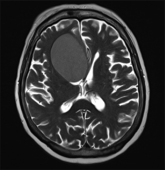
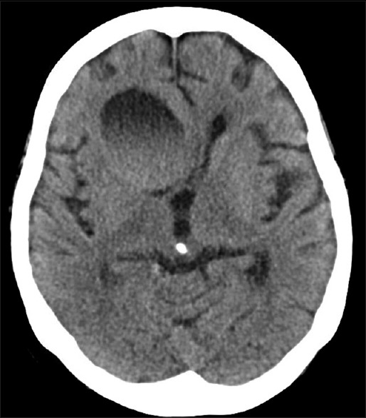
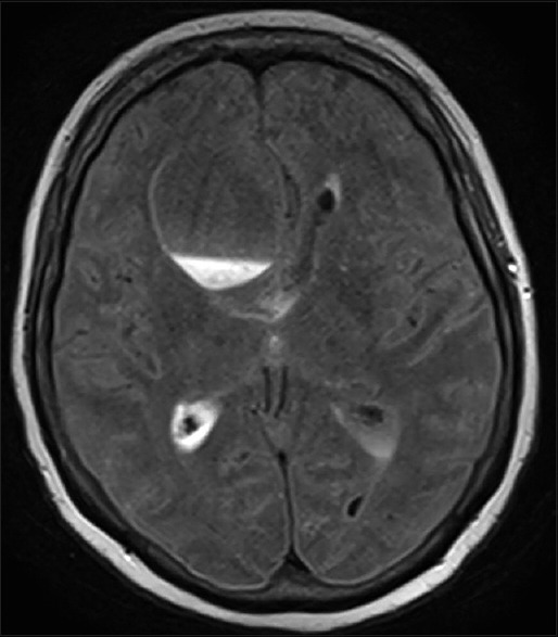
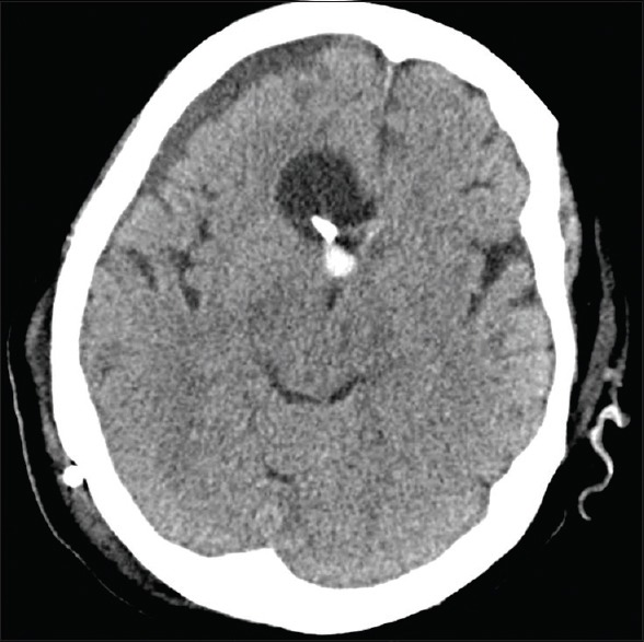
Intraventricular rupture of craniopharyngioma cysts is an unusual event which is associated with a high risk of loculated or communicating hydrocephalus. A 75-year-old woman presented at the Emergency Department of our hospital with mental status deterioration due to chemical ventriculitis and acute hydrocephalus following the intraventricular rupture of a craniopharyngioma cyst. The patient was treated with stress-dose steroid therapy. In addition, she underwent placement of an external ventricular drain and endoscopy-assisted intra-cystic placement of an Ommaya reservoir for the aspiration of the cystic fluid. The patient's condition improved; she was shunted in an expeditious fashion and discharged from the Intensive Care Unit within 2 weeks of her admission with the reservoir in place for the continued drainage of the cyst.
Keywords: Craniopharyngioma; cyst; hydrocephalus; intraventricular; rupture; supracellar; ventriculitis.
Conflict of interest statement
There are no conflicts of interest.
Figures
References
-
- Krueger DW, Larson EB. Recurrent fever of unknown origin, coma, and meningismus due to a leaking craniopharyngioma. Am J Med. 1988;84(3 Pt 1):543–5. - PubMed
-
- Maier HC. Craniopharyngioma with erosion and drainage into the nasopharynx. An autobiographical case report. J Neurosurg. 1985;62:132–4. - PubMed
-
- Okamoto H, Harada K, Uozumi T, Goishi J. Spontaneous rupture of a craniopharyngioma cyst. Surg Neurol. 1985;24:507–10. - PubMed
-
- Patrick BS, Smith RR, Bailey TO. Aseptic meningitis due to spontaneous rupture of craniopharyngioma cyst. Case report. J Neurosurg. 1974;41:387–90. - PubMed
-
- Ravindran M, Radhakrishnan VV, Rao VR. Communicating cystic craniopharyngioma. Surg Neurol. 1980;14:230–2. - PubMed
Publication types
LinkOut - more resources
Full Text Sources
Other Literature Sources