. 2017 Apr 20;7(8):e2234.
doi: 10.21769/BioProtoc.2234.

Expression, Purification and Crystallisation of the Adenosine A2A Receptor Bound to an Engineered Mini G Protein

Affiliations

Expression, Purification and Crystallisation of the Adenosine A2A Receptor Bound to an Engineered Mini G Protein

Byron Carpenter et al. Bio Protoc. .

Abstract

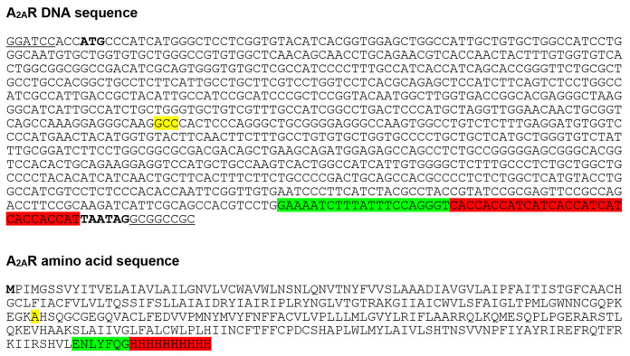
G protein-coupled receptors (GPCRs) promote cytoplasmic signalling by activating heterotrimeric G proteins in response to extracellular stimuli such as light, hormones and nucleosides. Structure determination of GPCR-G protein complexes is central to understanding the precise mechanism of signal transduction. However, these complexes are challenging targets for structural studies due to their conformationally dynamic and inherently transient nature. We recently developed an engineered G protein, mini-Gs, which addressed these problems and allowed the formation of a stable GPCR-G protein complex. Mini-Gs facilitated the structure determination of the human adenosine A2A receptor (A2AR) in its G protein-bound conformation at 3.4 Å resolution. Here, we describe a step by step protocol for the expression and purification of A2AR, and crystallisation of the A2AR-mini-Gs complex.

Keywords: A2AR; Active state; Adenosine A2A receptor; G protein complex; G protein-coupled receptor; GPCR; Mini G protein; Mini-Gs.


Figures

Figure 1.
Figure 1.. A2AR baculovirus expression construct.
A2AR was cloned into the baculovirus transfer vector pBacPAK8 using BamHI and NotI restriction sites (underlined), and baculoviruses were prepared using the flashBAC ULTRA system. The construct consists of residues 1-308 of human A2AR followed by a TEV protease cleavage site (highlighted in green) and C-terminal 10x histidine tag (highlighted in red). A2AR contains the N154A mutation (highlighted in yellow) to remove a potential N-linked glycosylation site. Start and stop codons are shown in bold.
Figure 2.
Figure 2.. Illustration of the membrane pellet after ultracentrifugation.
A diffuse layer, which contains a significant amount of A2AR, is observed on the surface of the membrane pellet after ultracentrifugation. It is important not to decant the supernatant, because this will remove the diffuse layer, resulting to a decreased yield of A2AR. Instead, the supernatant should be carefully aspirated using a serological pipette until level with the top of the membrane pellet (indicate by the dashed line).
Figure 3.
Figure 3.. SDS-PAGE and gel filtration analysis of the A2AR purification.
A and B. SDS-PAGE analysis of the purification of a representative A2AR construct. Despite having molecular weights of 35 and 27 kDa respectively, A2AR and mini-Gs414 migrate at an identical rate on the SDS-PAGE gel, making visualisation of the complex difficult. Therefore, for demonstration purposes, we show SDS-PAGE gels of an A2AR construct that contains an N-terminal thioredoxin fusion (TrxA-A2AR), which increases its molecular weight to 46 kDa. TrxA-A2AR was purified in DM using an identical protocol to that described for A2AR, however, the TrxA-A2AR–mini-Gs complex, which was exchanged into OTG, did not yield crystals that diffracted to higher than 3.5 Å resolution. A. SDS-PAGE analysis of the TrxA-A2AR purification: (M) molecular weight markers; (1) Ni2+-NTA column loading material (1:10 dilution); (2) Ni2+-NTA column flow through (1:10 dilution); (3) Ni2+-NTA column wash; (4) Ni2+-NTA column eluate; (5) sample after concentration and buffer exchange; (6) sample after TEV digestion; (7) Ni2+-NTA negative purification flow through; (8) Ni2+-NTA negative purification eluate; (9) gel filtration pool. TEV protease is indicated by an asterisk. The samples should not be boiled prior to loading on the gel (see Note 11). B. SDS-PAGE analysis of the TrxA-A2AR–mini-Gs414 complex after detergent exchange into OTG: (M) molecular weight markers; (1) gel filtration pool. C. Preparative gel filtration chromatogram for the A2AR–mini-Gs414 complex in OTG, pooled fractions (typically 2-3 ml) are indicated by dashed lines. Note that the UV detector reaches saturation, which results in a truncated absorbance profile. D. A crystal of the A2AR–mini-Gs414 complex mounted in a loop on beamline ID23-2 at the European Synchrotron Radiation Facility (the scale bar is 50 x 100 μm).

References

    1. Carpenter B., Nehmé R., Warne T., Leslie A. G. and Tate C. G.(2016). Structure of the adenosine A2A receptor bound to an engineered G protein . Nature 536(7614): 104-7. - PMC - PubMed
    1. Carpenter B. and Tate C. G.(2016). Engineering a minimal G protein to facilitate crystallisation of G protein-coupled receptors in their active conformation. Protein Eng Des Sel 29(12): 583-594. - PMC - PubMed
    1. Carpenter B. and Tate C. G.(2017). Expression and purification of mini G proteins from Escherichia coli . Bio-protocol 7(8): e2235. - PMC - PubMed
    1. Lebon G., Bennett K., Jazayeri A. and Tate C. G.(2011). Thermostabilisation of an agonist-bound conformation of the human adenosine A2A receptor . J Mol Biol 409(3): 298-310. - PMC - PubMed
    1. Lebon G., Warne T., Edwards P.C., Bennett K., Langmead C.J., Leslie A.G. and Tate C.G.(2011) Agonist-bound adenosine A2A receptor structures reveal common features of GPCR activation. Nature 474: 521-5. - PMC - PubMed