. 2017 Aug 15;196(4):479-493.
doi: 10.1164/rccm.201603-0568OC.

Differential Expression of VEGF-Axxx Isoforms Is Critical for Development of Pulmonary Fibrosis

Affiliations

Differential Expression of VEGF-Axxx Isoforms Is Critical for Development of Pulmonary Fibrosis

Shaney L Barratt et al. Am J Respir Crit Care Med. .

Abstract

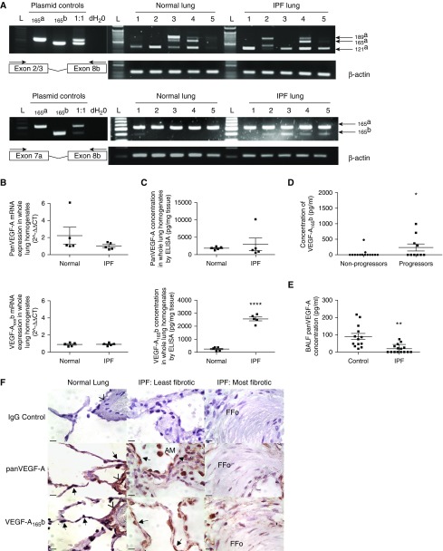
Rationale: Fibrosis after lung injury is related to poor outcome, and idiopathic pulmonary fibrosis (IPF) can be regarded as an exemplar. Vascular endothelial growth factor (VEGF)-A has been implicated in this context, but there are conflicting reports as to whether it is a contributory or protective factor. Differential splicing of the VEGF-A gene produces multiple functional isoforms including VEGF-A165a and VEGF-A165b, a member of the inhibitory family. To date there is no clear information on the role of VEGF-A in IPF.

Objectives: To establish VEGF-A isoform expression and functional effects in IPF.

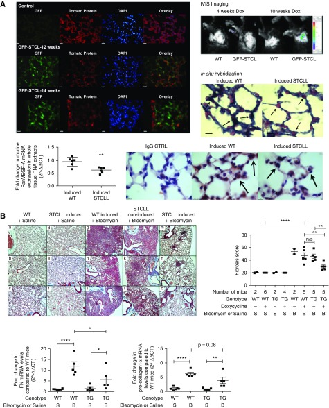
Methods: We used tissue sections, plasma, and lung fibroblasts from patients with IPF and control subjects. In a bleomycin-induced lung fibrosis model we used wild-type MMTV mice and a triple transgenic mouse SPC-rtTA+/-TetoCre+/-LoxP-VEGF-A+/+ to conditionally induce VEGF-A isoform deletion specifically in the alveolar type II (ATII) cells of adult mice.

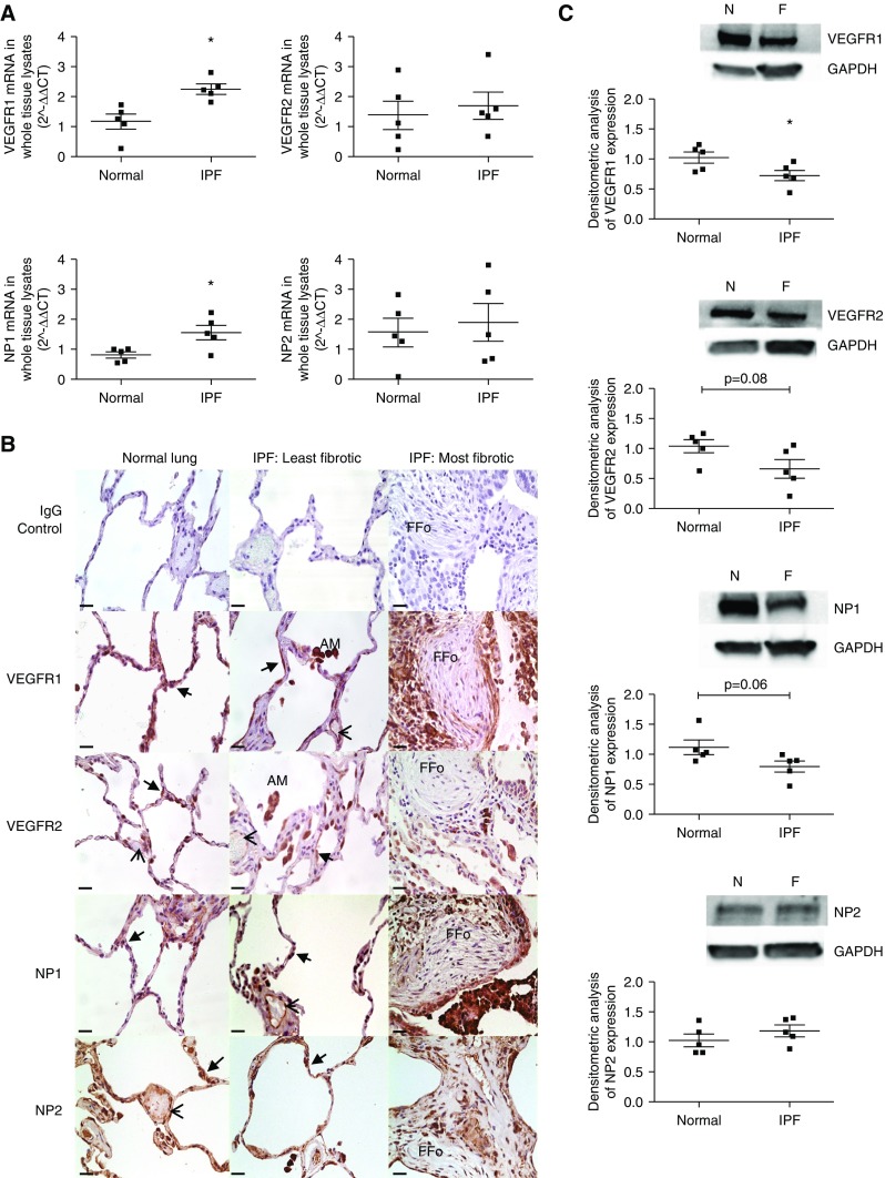
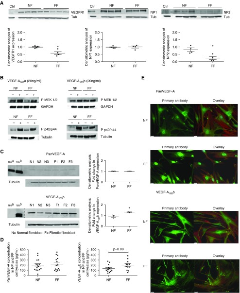
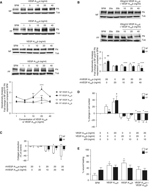
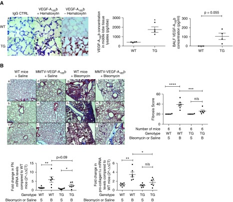
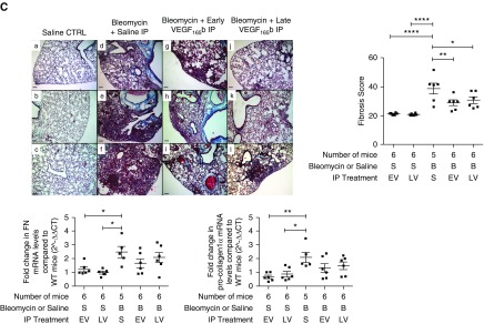
Measurements and main results: IPF and normal lung fibroblasts differentially expressed and responded to VEGF-A165a and VEGF-A165b in terms of proliferation and matrix expression. Increased VEGF-A165b was detected in plasma of progressing patients with IPF. In a mouse model of pulmonary fibrosis, ATII-specific deficiency of VEGF-A or constitutive overexpression of VEGF-A165b inhibited the development of pulmonary fibrosis, as did treatment with intraperitoneal delivery of VEGF-A165b to wild-type mice.

Conclusions: These results indicate that changes in the bioavailability of VEGF-A sourced from ATII cells, namely the ratio of VEGF-Axxxa to VEGF-Axxxb, are critical in development of pulmonary fibrosis and may be a paradigm for the regulation of tissue repair.

Keywords: animal models of pulmonary fibrosis; idiopathic pulmonary fibrosis; vascular endothelial growth factor.


Figures

Figure 1.
Figure 1.
Pan–vascular endothelial growth factor (VEGF)-A and VEGF-Axxxb expression in the lungs of patients with idiopathic pulmonary fibrosis (IPF). (A) Pan-VEGF-A reverse transcription–polymerase chain reaction (RT-PCR) of whole-lung RNA extract using VEGF-A Exon2/3 For and 8b Rev primers (top) and Exon 7a For and 8b Rev primers (bottom) (n = 5 normal lung; n = 5 IPF lung). VEGF-A121a, VEGF-A165a, VEGF-A165b, and VEGF-A189a isoforms were identified by RT-PCR and verified by direct sequencing (see Figure E1). L = 50-bp marker. (B) Quantitative RT-PCR of pan-VEGF-A and VEGF-Axxxb mRNA expression in whole-lung tissue homogenates of normal (n = 5) and IPF lung (n = 5). No significant difference was detected in the expression of pan-VEGF-A or VEGF-A165b (unpaired Student’s t test) isoforms between normal and IPF lung samples. (C) Pan-VEGF-A and VEGF-A165b ELISA data from lung whole-tissue lysates in normal (n = 5) and IPF (n = 5) subjects. There was no significant difference in pan-VEGF-A expression (unpaired Student’s t test with Welch correction), but a significant increase in VEGF-A165b expression in the IPF lung was observed (****P < 0.0001; unpaired Student’s t test). Data are presented as means with SEM. (D) VEGF-A165b ELISA data from plasma samples of progressor (death or >10% decline in FVC at 12 mo follow-up) and nonprogressor patients with IPF (38). There was a significant difference in plasma levels of VEGF-A165b in progressors (n = 10) compared with nonprogressors (n = 15) (*P < 0.05; unpaired Student’s t test). (E) Pan-VEGF-A levels in bronchoalveolar lavage fluid (BALF) of patients with IPF (n = 15) compared with control subjects (n = 13) using an antibody that does not discriminate between isoforms. Patient demographics were statistically comparable by unpaired Student’s t test (see Figure E2A). Pan-VEGF-A BALF levels were significantly lower in the IPF group compared with control subjects, **P < 0.01, unpaired Student’s t test with Welch correction. Data are presented as means with SEM. Mean pan-VEGF-A expression in control group 85.7 pg/ml ± 17.1, versus IPF 18.0 pg/ml ±  6.1. VEGF-A165b expression was below the limit of detection in both patients with IPF and control subjects (10 pg/ml). (F) Pan-VEGF-A and VEGF-A165b immunohistochemical staining. Intense staining of the alveolar epithelium was observed for both pan-VEGF-A and VEGF-A165b in normal and IPF lungs (least and most fibrotic designated as in Ashcroft and colleagues [59] and Ebina and colleagues [60]) (arrows). Additional sites of localization included vessel walls (arrowheads), fibroblasts, lymphocytes, and alveolar macrophages. Isotype IgG shown as negative control. Scale bars = 10 μm; original magnification, ×100. Lower-magnification images are shown in Figure E2B. AM = alveolar macrophages; FFo = fibrotic focus.
Figure 2.
Figure 2.
Vascular endothelial growth factor (VEGF)-A receptor and coreceptor expression in the lungs of patients with idiopathic pulmonary fibrosis (IPF). (A) Quantitative reverse transcription–polymerase chain reaction of VEGFR1, VEGFR2, neuropilin (NP) 1, and NP2 mRNA expression in whole-lung RNA extracts of normal (n = 5) and IPF (n = 5) lung. VEGFR1 (*P < 0.05; unpaired Student’s t test) and NP1 (*P < 0.05; unpaired Student’s t test) mRNA expression was significantly up-regulated in the IPF lung. Data are presented as mean fold change in expression (2−ΔΔCT) with SEM, data analysis performed on ΔΔCT values. (B) VEGF-A receptor and coreceptor immunohistochemical staining. Intense staining of the alveolar epithelium was observed for VEGFR1, VEGFR2, NP1, and NP2 (arrows) in both the normal and IPF lung. Additional sites of localization included the vascular endothelium (arrowheads), fibroblasts, lymphocytes, and alveolar macrophages. Isotype IgG shown as negative control subject. Images were taken at ×40 magnification; scale bars = 25 μm. Additional higher-magnification images are shown in Figure E3B. (C) Expression of VEGFR1, VEGFR2, NP1, and NP2 in whole-tissue lysates of normal (n = 5) and IPF (n = 5) lung by Western blotting (top) with semiquantitative densitometric analysis (bottom). VEGFR1, VEGFR2, NP1, and NP2 were expressed in both the normal and IPF lung. VEGFR1 (*P < 0.05) was significantly down-regulated in the IPF lung (unpaired Student’s t test, data presented as mean densitometry score with SEM). AM = alveolar macrophages; F = IPF lung; FFo = fibrotic focus; N = normal lung.
Figure 3.
Figure 3.
Vascular endothelial growth factor receptor (VEGFR) and vascular endothelial growth factor (VEGF)-A isoform expression in NF and FF. (A) Expression of VEGFR1, VEGFR2, neuropilin (NP) 1, and NP2 in NF and FF by Western blotting with semiquantitative densitometric analysis. Using glomerular endothelial cells as a positive control (Ctrl), bands were observed by Western blotting that were consistent with the expression of VEGFR1, NP1, and NP2 in both NF and FF. Mature VEGFR2 protein was not expressed by NF or FF fibroblasts (data not shown). Semiquantification of expression by densitometry demonstrated a significant reduction in the expression of VEGFR1 (*P < 0.05) and NP2 (*P < 0.05) in unstimulated FF cultures compared with NF (data presented as means with SEM; NF, n = 4; FF, n = 4, n = 3 or 4 shown; unpaired Student’s t test). (B) Western blot of phosphorylated MEK1/2 and phosphorylated p42/p44 MAPK (mitogen-activated protein kinase) expression in response to 24 hours of stimulation with VEGF-A165a (20 ng/ml) or VEGF-A165b (20 ng/ml). In the absence of mature VEGFR2 expression in NF and FF, the activation of known VEGF-A signaling pathways was explored. Stimulation of NF and FF led to the increased phosphorylation of MEK1/2 and p42/p44 in response to treatment of cells with VEGF-A165a or VEGF-A165b. For densitometric analysis see Figure E4. (C) Western blot of pan-VEGF-A and VEGF-A165b expression in NF and FF cell lysates with semiquantitative densitometric analysis (data presented as means with SEM). Recombinant proteins were used as positive control subjects to highlight the specificity of the VEGF-A165b antibody in detecting VEGF-A165b proteins. The dashed line in the lower blot indicates where the blot has been manually cut and components imaged separately. VEGF-A165b proteins were significantly up-regulated in FF cell lysates compared with NF (n = 3; *P < 0.05; unpaired Student’s t test). No significant difference in pan-VEGF-A isoform expression was shown (n = 3; unpaired Student’s t test). (D) Quantification of protein expression of pan-VEGF-A and VEGF-A165b expression in NF and FF cell lysates. By ELISA there was no significant difference in the expression of pan-VEGF-A or VEGF-A165b in NF or FF cell lysates (n = 6 performed in duplicate cell lysates of different passage, unpaired Student’s t test). In the conditioned medium extracted from these cultures pan-VEGF-A expression was significantly up-regulated in the FF supernatants (n = 6; *P < 0.05; unpaired Student’s t test) (see Figure E7). VEGF-A165b expression in these same cell supernatant samples was below the limit of detection of the ELISA (10 pg/ml). (E) Cell immunofluorescence of pan-VEGF-A and VEGF-A165b expression in NF and FF. Comparable patterns were observed for NF and FF, with cytoplasmic and perinuclear expression of pan-VEGF-A and cytoplasmic expression of VEGF-A165b. Images were taken at ×40 magnification with scale bar = 25 μm. Primary antibody shown in green with an overlay image of the primary antibody, phalloidin (red) for F-actin and DAPI (blue) for nuclear staining. Isotype IgG control subjects and separate phalloidin and DAPI images shown in Figure E8. FF = patients with idiopathic pulmonary fibrosis; NF = normal subjects; Tub = tubulin.
Figure 4.
Figure 4.
Functional response of normal subjects (NF) and patients with idiopathic pulmonary fibrosis (FF) to recombinant vascular endothelial growth factor (VEGF)-A proteins. (A) The effect of VEGF-A on the expression of fibronectin (FN) by NF and FF in response to 24 hours of stimulation with VEGF-A165a (20 ng/ml) or VEGF-A165b (20 ng/ml) as measured by Western blotting (top) with densitometry analysis (bottom). A significant increase in the expression of FN in response to VEGF-A165a stimulation in both NF (**P < 0.01 with 10 ng/ml and 20 ng/ml) and FF (+++P < 0.001 at 20 ng/ml; n = 3) was observed. There was no significant effect of VEGF-A165b on the expression of FN in either NF or FF. (B) The combined effect of VEGF-A165a and VEGF-A165b on the expression of FN in NF and FF as measured by Western blotting (top) and densitometry (bottom). Used in isolation, VEGF-A165a (20 ng/ml) was shown to increase FN expression in NF (XP < 0.05) and FF (XXP < 0.01), whereas VEGF-A165b had no significant effect, as shown in the previous experiments. VEGF-A165b inhibited the VEGF-A165a–induced increase in FN expression in both NF (*P < 0.05 at 40 ng/ml of VEGF-A165b) and FF (+++P < 0.001 at 10 and 40 ng/ml of VEGF-A165b, ++P < 0.01 at 20 ng/ml of VEGF-A165b), with FF appearing to be more susceptible to this effect (n = 3). Data are presented as means with SEM. (C) The effect of VEGF-A on the expression of collagen by NF and FF in response to 24 hours of stimulation with VEGF-A165a or VEGF-A165b as measured by HPLC. In the presence of both isoforms, VEGF-A165b inhibited collagen production in NF but not FF (*P < 0.05; **P < 0.01; NF and FF n = 3). Data are presented as means with SEM. (D) The effect of VEGF-A recombinant proteins on the proliferation of NF and FF. A significant increase in FF cell number was observed in response to 20 ng/ml VEGF-A165a compared with SFM (XP < 0.05). VEGF-A165a-induced proliferation was inhibited by the concomitant addition of 20 ng/ml VEGF-A165b (+++P < 0.001) or 10 ng/ml sFlt (+++P < 0.001). VEGF-A165a had no statistically significant effect on NF cell proliferation compared with SFM, but concomitant treatment of NF with 20 ng/ml VEGF-A165a and 20 ng/ml VEGF-A165b (*P < 0.05) or sFlt (*P < 0.05) inhibited cell proliferation compared with 20 ng/ml VEGF-A165a alone (*P < 0.05; NF and FF n = 5). Data are presented as means with SEM. (E) The effect of VEGF-A recombinant proteins on NF and FF wound healing. Image analysis performed using ImageJ. VEGF-A165a and VEGF-A165b significantly increased the migration of NF at 48 hours (*P < 0.05). This effect was blocked by the concomitant treatment of VEGF-A165a and VEGF-A165b. Recombinant VEGF-A proteins had no significant effect on the migration of FF. Statistical analysis: analysis of variance with post hoc Holm-Šidák multiple comparisons analysis used throughout (n = 4). Representative images shown in Figure E9. SFM = serum-free media; Tub = tubulin.
Figure 5.
Figure 5.
The effect of postnatal deletion of vascular endothelial growth factor (VEGF)-A from alveolar type II (ATII) cells on the development of pulmonary fibrosis. (A) GFP-reporter mice (ROSAmT/mG) were crossed with SPC+/−TC+/−LoxP+/− transgenic (TG) mice (STCL) to determine the period of doxycycline induction required to activate the Cre-recombinase and to identify the tissue specificity of Cre-recombinase activity after doxycycline induction. The reporter mice possess LoxP sites on either side of a membrane-targeted tdTomato (mT) cassette and express red fluorescence in all tissues. In the presence of Cre recombinase, the mT cassette is deleted, allowing expression of the membrane-targeted EGFP (mG) cassette located just downstream. The in vivo imaging system (IVIS) demonstrated evidence of GFP fluorescence in the GFP-STCL mouse after 10 weeks of doxycycline induction, absent at 4 weeks of induction and in wild-type (WT) mice. Direct visualization of the lungs of induced mice (at 12 and 14 wk of doxycycline treatment) demonstrated GFP fluorescence in ATII cells with an absence of GFP fluorescence in the bronchial epithelium (14-wk induction image; arrows). Images were taken at ×40 magnification; scale bar = 25 μm, n = 3. GFP fluorescence was absent in the kidneys and liver (see Figure E10). Using a murine VEGF-A RNA probe, in situ hybridization demonstrated a reduction in ATII cell staining (reduction in blue/black signal) in induced-TG mice compared with induced-WT mice (arrows). Pink staining represents nuclear fast red counterstain (n = 3; images taken at ×100 magnification; scale bar = 10 μm). Pan-VEGF-A mRNA expression was significantly reduced in whole-tissue RNA extracts from TG mice compared with WT mice (n = 6; **P < 0.01; unpaired Student’s t test; data presented as means with SEM). Consistent with these findings, immunohistochemical staining for pan-VEGF-A also showed a reduction in ATII cell staining in induced-TG mice compared with induced-WT mice (n = 3; images taken at ×100 magnification; scale bar = 10 μm). (B) Postnatal deletion of VEGF-A from ATII cells ameliorates the development of bleomycin (BLM)-induced pulmonary fibrosis. Masson trichrome staining of mouse lung sections, with lung fibrosis score (above) and quantitative reverse transcription–polymerase chain reaction of FN and procollagen-1α mRNA levels (below). As demonstrated by Masson trichrome staining, administration of BLM to both WT (g–i) and noninduced STCLL (j–l) mice resulted in extensive pulmonary fibrosis. This effect was ameliorated in BLM-treated doxycycline-induced STCLL mice (m–o). Images were originally ×10 magnification; scale bar = 100 μm; n = 5 or 6; n = 3 shown. The lung fibrosis score of BLM-treated, doxycycline-induced STCLL mice was significantly reduced compared with BLM-treated control animals (induced-WT mice **P < 0.01 and noninduced STCLL mice **P < 0.01; n = 5). The expression of FN and pro-collagen-1α mRNA was significantly up-regulated in the lungs of BLM-treated, doxycycline-induced WT mice compared with saline-treated control animals (****P < 0.001; n = 5 or 6 analyzed). FN and pro-collagen-1α expression was significantly up-regulated in BLM-treated, doxycycline-induced TG mice compared with TG saline controls (*P < 0.05; **P < 0.01; n = 5). Furthermore, FN mRNA levels were significantly reduced in BLM-treated TG mice compared with BLM-treated WT mice (*P < 0.05; n = 5), with a trend toward a reduction in pro-collagen-1α levels in these same mice (P = 0.08, n = 5). Statistical analysis: analysis of variance with Holm-Šidák multiple comparisons test used throughout. n/s = not significant; STCLL = SPC-rtTA+/−TetoCre+/−LoxP-VEGF-A+/+.
Figure 6.
Figure 6.
The effect of vascular endothelial growth factor (VEGF)-A165b on the development of pulmonary fibrosis. (A) Lung phenotype of the MMTV-VEGF-A165b transgenic (TG) mouse. Immunohistochemical staining for VEGF-A165b in the lung of TG mice with quantification of VEGF-A165b expression by ELISA, in whole-tissue lysates and bronchoalveolar lavage fluid (BALF). VEGF-A165b expression was increased in the alveolar type II (ATII) cells of the TG mouse lung (d and f, indicated by arrows) compared with the wild-type (WT) lung (c and e). Isotype IgG staining was used as a negative control (a and b). Images were taken at ×100 magnification; scale bars = 10 μm, (n = 3, n = 1 shown). VEGF-A165b expression was significantly up-regulated in whole-tissue lysates of TG mice compared with WT mice (*P < 0.05; unpaired Student’s t test with Welch correction; n = 4). The expression of VEGF-A165b was below the limit of detection of the ELISA in all BALF samples of WT mice but present in detectable levels in all BALF samples of TG mice. There was no statistical difference between these groups (P = 0.055; unpaired Student’s t test with Welch correction; n = 4). (B) Overexpression of VEGF-A165b in ATII cells ameliorates the development of bleomycin (BLM)-induced pulmonary fibrosis. Representative images of Masson trichrome staining with lung fibrosis score and quantitative reverse transcription–polymerase chain reaction (RT-PCR) of fibronectin (FN) and pro-collagen-1α mRNA levels (bottom). Masson trichrome staining of mouse lung sections, 21 days after oropharyngeal (OP) instillation of BLM to MMTV-VEGF-A165b TG mice (j–l) or littermate control animals (g–i). The development of BLM-induced pulmonary fibrosis was ameliorated in TG mice. Saline control subjects are shown in a–f (n = 6 per group; n = 3 shown). Scale bar = 100 μm; original magnification, ×10. The lung fibrosis score of BLM-treated WT mice was significantly greater than saline-treated WT mice (****P < 0.0001; n = 6 per group). The lung fibrosis score of BLM-treated TG mice was significantly lower than BLM-treated WT mice (***P < 0.001; n = 6 per group). FN mRNA was significantly up-regulated in the lungs of WT BLM-treated mice compared with WT saline-treated control animals (**P < 0.01; n = 6 analyzed) and in TG BLM-treated mice compared with TG saline-treated control animals (*P < 0.05; n = 6). There was no statistical difference between BLM-treated WT and TG mice (P = 0.09; n = 6). In contrast, pro-collagen-1α mRNA levels were significantly reduced in BLM-treated TG mice compared with BLM-treated WT littermates (*P < 0.05; n = 6). Data are presented as means with SEM. Statistical analysis: analysis of variance with Holm-Šidák multiple comparisons test used throughout. (C) IP instillation of rhVEGF-A165b ameliorates the development of pulmonary fibrosis. Masson trichrome staining of lung sections with lung fibrosis score and quantitative RT-PCR of FN and pro-collagen-1α mRNA levels (bottom). Representative images of Masson trichrome staining of mouse lung sections, 21 days after OP instillation of BLM with or without additional IP rhVEGF-A165b instillation. (a–c) OP saline control (no differences between early IP VEGF-A165b [EV] or late IP VEGF-A165b [LV] plus saline). (d–f) OP BLM with saline IP. (g–i) Early IP VEGF-A165b treatment with subsequent BLM OP instillation. (j–l) BLM OP with subsequent late IP VEGF-A165b instillation. Early and late rhVEGF-A165b instillation ameliorated the development of lung fibrosis (n = 6 per group; n = 3 shown). Scale bar = 100 μm; original magnification, ×10. By qRT-PCR, FN and pro-collagen-1α mRNA was significantly up-regulated in the lungs of BLM-treated mice compared with saline-treated control animals (*P < 0.05; **P < 0.01; n = 5). In contrast, FN and pro-collagen-1α expression in mice treated with either early (EV) or late (LV) rhVEGF-A165b and BLM was not significantly different from saline control (n = 6). The lung fibrosis score in both early (EV, **P < 0.01) and late (LV, *P < 0.05) IP rhVEGF-A165b treatment groups was significantly reduced compared with BLM OP–saline IP–treated mice; n = 5 or 6 per group; data are presented as means with SEM. Statistical analysis: analysis of variance with Holm-Šidák multiple comparisons test used throughout. CTRL = control; n/s = not significant.
Figure 6.
Figure 6.
The effect of vascular endothelial growth factor (VEGF)-A165b on the development of pulmonary fibrosis. (A) Lung phenotype of the MMTV-VEGF-A165b transgenic (TG) mouse. Immunohistochemical staining for VEGF-A165b in the lung of TG mice with quantification of VEGF-A165b expression by ELISA, in whole-tissue lysates and bronchoalveolar lavage fluid (BALF). VEGF-A165b expression was increased in the alveolar type II (ATII) cells of the TG mouse lung (d and f, indicated by arrows) compared with the wild-type (WT) lung (c and e). Isotype IgG staining was used as a negative control (a and b). Images were taken at ×100 magnification; scale bars = 10 μm, (n = 3, n = 1 shown). VEGF-A165b expression was significantly up-regulated in whole-tissue lysates of TG mice compared with WT mice (*P < 0.05; unpaired Student’s t test with Welch correction; n = 4). The expression of VEGF-A165b was below the limit of detection of the ELISA in all BALF samples of WT mice but present in detectable levels in all BALF samples of TG mice. There was no statistical difference between these groups (P = 0.055; unpaired Student’s t test with Welch correction; n = 4). (B) Overexpression of VEGF-A165b in ATII cells ameliorates the development of bleomycin (BLM)-induced pulmonary fibrosis. Representative images of Masson trichrome staining with lung fibrosis score and quantitative reverse transcription–polymerase chain reaction (RT-PCR) of fibronectin (FN) and pro-collagen-1α mRNA levels (bottom). Masson trichrome staining of mouse lung sections, 21 days after oropharyngeal (OP) instillation of BLM to MMTV-VEGF-A165b TG mice (j–l) or littermate control animals (g–i). The development of BLM-induced pulmonary fibrosis was ameliorated in TG mice. Saline control subjects are shown in a–f (n = 6 per group; n = 3 shown). Scale bar = 100 μm; original magnification, ×10. The lung fibrosis score of BLM-treated WT mice was significantly greater than saline-treated WT mice (****P < 0.0001; n = 6 per group). The lung fibrosis score of BLM-treated TG mice was significantly lower than BLM-treated WT mice (***P < 0.001; n = 6 per group). FN mRNA was significantly up-regulated in the lungs of WT BLM-treated mice compared with WT saline-treated control animals (**P < 0.01; n = 6 analyzed) and in TG BLM-treated mice compared with TG saline-treated control animals (*P < 0.05; n = 6). There was no statistical difference between BLM-treated WT and TG mice (P = 0.09; n = 6). In contrast, pro-collagen-1α mRNA levels were significantly reduced in BLM-treated TG mice compared with BLM-treated WT littermates (*P < 0.05; n = 6). Data are presented as means with SEM. Statistical analysis: analysis of variance with Holm-Šidák multiple comparisons test used throughout. (C) IP instillation of rhVEGF-A165b ameliorates the development of pulmonary fibrosis. Masson trichrome staining of lung sections with lung fibrosis score and quantitative RT-PCR of FN and pro-collagen-1α mRNA levels (bottom). Representative images of Masson trichrome staining of mouse lung sections, 21 days after OP instillation of BLM with or without additional IP rhVEGF-A165b instillation. (a–c) OP saline control (no differences between early IP VEGF-A165b [EV] or late IP VEGF-A165b [LV] plus saline). (d–f) OP BLM with saline IP. (g–i) Early IP VEGF-A165b treatment with subsequent BLM OP instillation. (j–l) BLM OP with subsequent late IP VEGF-A165b instillation. Early and late rhVEGF-A165b instillation ameliorated the development of lung fibrosis (n = 6 per group; n = 3 shown). Scale bar = 100 μm; original magnification, ×10. By qRT-PCR, FN and pro-collagen-1α mRNA was significantly up-regulated in the lungs of BLM-treated mice compared with saline-treated control animals (*P < 0.05; **P < 0.01; n = 5). In contrast, FN and pro-collagen-1α expression in mice treated with either early (EV) or late (LV) rhVEGF-A165b and BLM was not significantly different from saline control (n = 6). The lung fibrosis score in both early (EV, **P < 0.01) and late (LV, *P < 0.05) IP rhVEGF-A165b treatment groups was significantly reduced compared with BLM OP–saline IP–treated mice; n = 5 or 6 per group; data are presented as means with SEM. Statistical analysis: analysis of variance with Holm-Šidák multiple comparisons test used throughout. CTRL = control; n/s = not significant.

Comment in

References

    1. Navaratnam V, Fleming KM, West J, Smith CJ, Jenkins RG, Fogarty A, Hubbard RB. The rising incidence of idiopathic pulmonary fibrosis in the U.K. Thorax. 2011;66:462–467. - PubMed
    1. Collard HR, King TE, Jr, Bartelson BB, Vourlekis JS, Schwarz MI, Brown KK. Changes in clinical and physiologic variables predict survival in idiopathic pulmonary fibrosis. Am J Respir Crit Care Med. 2003;168:538–542. - PubMed
    1. Raghu G, Weycker D, Edelsberg J, Bradford WZ, Oster G. Incidence and prevalence of idiopathic pulmonary fibrosis. Am J Respir Crit Care Med. 2006;174:810–816. - PubMed
    1. Olson AL, Swigris JJ, Lezotte DC, Norris JM, Wilson CG, Brown KK. Mortality from pulmonary fibrosis increased in the United States from 1992 to 2003. Am J Respir Crit Care Med. 2007;176:277–284. - PubMed
    1. Ferrara N. Vascular endothelial growth factor: basic science and clinical progress. Endocr Rev. 2004;25:581–611. - PubMed

Publication types

Substances