. 2017 Jul;6(7):1644-1659.
doi: 10.1002/sctm.16-0222. Epub 2017 Jun 8.

Endochondral Ossification in Critical-Sized Bone Defects via Readily Implantable Scaffold-Free Stem Cell Constructs

Affiliations

Endochondral Ossification in Critical-Sized Bone Defects via Readily Implantable Scaffold-Free Stem Cell Constructs

Phuong N Dang et al. Stem Cells Transl Med. 2017 Jul.

Abstract

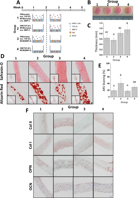
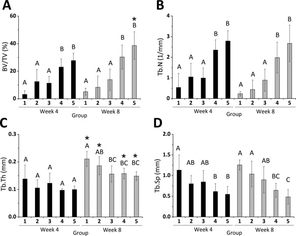
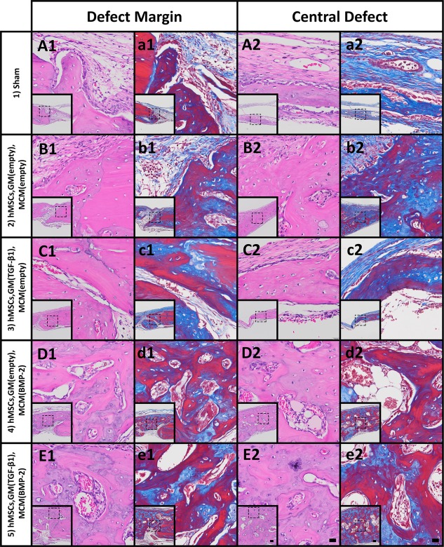
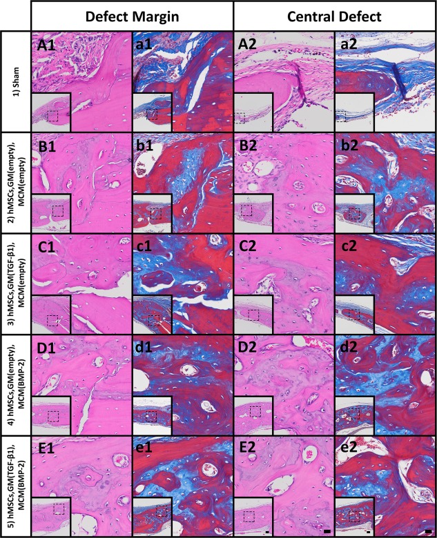
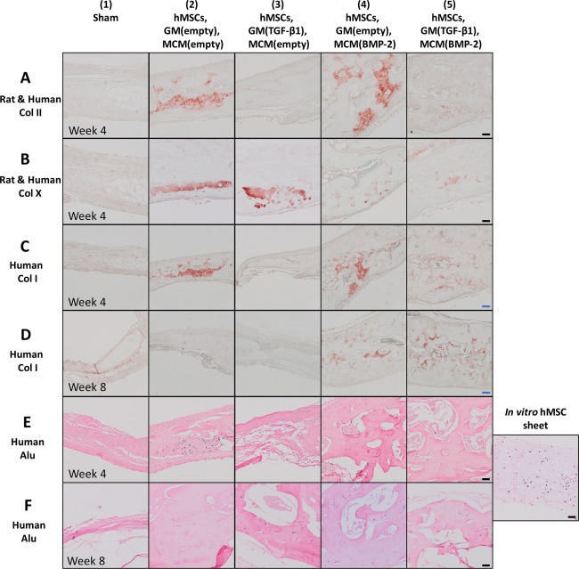
The growing socioeconomic burden of musculoskeletal injuries and limitations of current therapies have motivated tissue engineering approaches to generate functional tissues to aid in defect healing. A readily implantable scaffold-free system comprised of human bone marrow-derived mesenchymal stem cells embedded with bioactive microparticles capable of controlled delivery of transforming growth factor-beta 1 (TGF-β1) and bone morphogenetic protein-2 (BMP-2) was engineered to guide endochondral bone formation. The microparticles were formulated to release TGF-β1 early to induce cartilage formation and BMP-2 in a more sustained manner to promote remodeling into bone. Cell constructs containing microparticles, empty or loaded with one or both growth factors, were implanted into rat critical-sized calvarial defects. Micro-computed tomography and histological analyses after 4 weeks showed that microparticle-incorporated constructs with or without growth factor promoted greater bone formation compared to sham controls, with the greatest degree of healing with bony bridging resulting from constructs loaded with BMP-2 and TGF-β1. Importantly, bone volume fraction increased significantly from 4 to 8 weeks in defects treated with both growth factors. Immunohistochemistry revealed the presence of types I, II, and X collagen, suggesting defect healing via endochondral ossification in all experimental groups. The presence of vascularized red bone marrow provided strong evidence for the ability of these constructs to stimulate angiogenesis. This system has great translational potential as a readily implantable combination therapy that can initiate and accelerate endochondral ossification in vivo. Importantly, construct implantation does not require prior lengthy in vitro culture for chondrogenic cell priming with growth factors that is necessary for current scaffold-free combination therapies. Stem Cells Translational Medicine 2017;6:1644-1659.

Keywords: Adult stem cells; Bone tissue engineering; Growth factor delivery; Microparticles; Regenerative medicine; Self-assembled sheets.


Figures

Figure 1
Figure 1
Analysis of scaffold‐free hMSC constructs cultured in vitro for 5 weeks. (A): Schematic of conditions. Constructs were incorporated with both GM and MCM that were either left empty or loaded with TGF‐β1 or BMP‐2, respectively. Each group was cultured for 2 weeks in serum‐free basal medium with or without exogenously supplemented (exo.) TGF‐β1 and then for 3 weeks in serum‐free osteogenic medium with or without exo. BMP‐2. (B): Gross images and (C): thickness measurement of constructs after 5 weeks of in vitro culture. Groups that do not share a letter are significantly different at p < .05. (D): Photomicrographs of representative Safranin O and ARS stained sections of constructs after 5 weeks of culture. Scale bars = 100 μm, 500 μm (inset). All images are at the same scale. (E): Quantitative analysis of ARS staining. Groups that do not share a letter are significantly different at p < .05. (F): Photomicrographs of representative Col II, Col I, OPN, and OCN sections of microparticle‐incorporated sheets after 5 weeks of in vitro culture. Scale bar = 100 μm. All images are at the same scale. Abbreviations: ARS, alizarin red S; BMP‐2, bone morphogenetic protein‐2; Col I, type I collagen; Col II, type II collagen; GM, gelatin microparticles; hMSC, human mesenchymal stem cell; MCM, mineral‐coated hydroxyapatite microparticles; OCN, osteocalcin; OPN, osteopontin; TGF‐β1, transforming growth factor‐beta 1.
Figure 2
Figure 2
In vivo study conditions and real‐time PCR analysis of growth factor receptor expression and cartilage and bone marker expression in day 2 constructs prior to implantation. (A): Schematic of in vivo study conditions. (B–G): Real‐time PCR analysis of mRNA expression of (B) TGFBR1, (C) TGFBRII, (D) VEGFRII, (E) BMPRIA, (F) BMPRIB, and (G) BMPRII. (H–M): Real‐time PCR analysis of mRNA expression of early and late cartilage markers (H) SOX9, (I) ACAN, (J) COL2A1 and bone markers (K) RUNX2, (L) ALP, and (M) OCN. Groups that do not share a letter are significantly different at p < .05. Abbreviations: ACAN, aggrecan; ALP, alkaline phosphatase; BMP‐2, bone morphogenetic protein‐2; BMPRIA, BMP‐2 receptor type IA; BMPRIB, BMP‐2 receptor type IB; BMPRII, BMP‐2 receptor type II; COL2A1, collagen type 2A1; GM, gelatin microparticles; hMSC, human mesenchymal stem cell; OCN, osteocalcin; RUNX2, Runt‐related transcription factor 2; SOX9, sex determining Y‐box 9; TGF‐β1, transforming growth factor‐beta 1; TGFBR1, TGF‐β1 receptor type I; TGFBRII, TGF‐β1 receptor type II; VEGFRII, vascular endothelial growth factor receptor type II.
Figure 3
Figure 3
Micro‐CT reconstructions of defect regions after 4 and 8 weeks post‐implantation and micro‐CT scoring for bony bridging and union within defects. (A): Three‐dimensional reconstructed micro‐CT images (top) and Tb.Th maps (bottom) of the defect region in representative samples for each time point. Tb.Th increased from blue to green to red. White scale bar = 500 μm. All images are at the same scale. (B): Scoring guide for extent of bony bridging and union using micro‐CT reconstructions (adapted from Patel et al. 47). (C): Scores for bony bridging and union for the five in vivo groups examined at 4 and 8 weeks. Groups that do not share a letter are significantly different at p < .05. Abbreviations: micro‐CT, micro‐computed tomography; Tb.Th, trabecular thickness.
Figure 4
Figure 4
Quantitative micro‐computed tomography (micro‐CT) analysis. (A) BV/TV, (B) Tb.N, (C) Tb.Th, and (D) Tb.Sp of newly formed bone in the defect region at 4 and 8 weeks from micro‐CT data. Within each time point, groups that do not share a letter are significantly different at p < .05. * Signifies statistical significance compared to week 4. Abbreviations: BV/TV, bone volume/total volume; Tb.N, trabecular number; Tb.Sp, trabecular separation; Tb.Th, trabecular thickness.
Figure 5
Figure 5
Histological evaluation after 4 weeks. Photomicrographs of (A1–E1): H&E‐ and (a1–e1): Masson's trichrome‐stained sections at the defect margin. Photomicrographs of (A2–E2): H&E‐ and (a2–e2): Masson's trichrome‐stained sections at a central region of the defect. Scale bars = 100 µm (inset), 20 µm (magnification of dotted squares). Abbreviations: BMP‐2, bone morphogenetic protein‐2; GM, gelatin microparticles; hMSC, human mesenchymal stem cell; MCM, mineral‐coated hydroxyapatite microparticles; TGF‐β1, transforming growth factor‐beta 1.
Figure 6
Figure 6
Histological evaluation after 8 weeks. Photomicrographs of (A1–E1): H&E‐ and (a1–e1): Masson's trichrome‐stained sections at the defect margin. Photomicrographs of (A2–E2): H&E‐ and (a2–e2): Masson's trichrome‐stained sections at a central region of the defect. Scale bars = 100 µm (inset), 20 µm (magnification of dotted squares). Abbreviations: BMP‐2, bone morphogenetic protein‐2; GM, gelatin microparticles; hMSC, human mesenchymal stem cell; MCM, mineral‐coated hydroxyapatite microparticles; TGF‐β1, transforming growth factor‐beta 1.
Figure 7
Figure 7
Immunohistological evaluation for extracellular matrix components present in cartilage and bone in the rat calvarial defects at weeks 4 and 8. Photomicrographs of sections at the central defect region stained for (A): human and rat Col II at week 4, (B): human and rat Col X at week 4, (C): human Col I at week 4, and (D): human Col I at week 8. Photomicrographs of human Alu stained sections at the central defect region at (E): week 4 and (F): week 8 and of human Alu staining positive control, a scaffold‐free hMSC construct cultured for 5 weeks in vitro. Scale bars = 50 μm (black), 100 μm (blue). Abbreviations: BMP‐2, bone morphogenetic protein‐2; Col X, collagen X; Col I, type I collagen; Col II, type II collagen; GM, gelatin microparticles; hMSC, human mesenchymal stem cell; MCM, mineral‐coated hydroxyapatite microparticles; TGF‐β1, transforming growth factor‐beta 1.

References

    1. Meijer GJ, de Bruijn JD, Koole R et al. Cell‐based bone tissue engineering. PLoS Med 2007;4:e9. - PMC - PubMed
    1. Chen SS, Fitzgerald W, Zimmerberg J et al. Cell‐cell and cell‐extracellular matrix interactions regulate embryonic stem cell differentiation. Stem Cells 2007;25:553–561. - PubMed
    1. Levy‐Mishali M, Zoldan J, Levenberg S. Effect of scaffold stiffness on myoblast differentiation. Tissue Eng Part A 2009;15:935–944. - PubMed
    1. Reis RL, Cohn D. Polymer based systems on tissue engineering, replacement and regeneration. NATO Science Series, Series II: Mathematics, Physics and Chemistry, 1568‐2609 86. Dordrecht: Springer Netherlands: Imprint: Springer, 2002:1 online resource (440 pages).
    1. Langer RS, Lanza RP, Vacanti J. Principles of Tissue Engineering: Elsevier Inc., 2014: 1 online resource (xlviii, 1887 pages). https://www.elsevier.com/books/principles-of-tissue-engineering/lanza/97...

Publication types