

Review
doi: 10.1164/rccm.201703-0451PP.
The Role of Chest Computed Tomography in the Evaluation and Management of the Patient with Chronic Obstructive Pulmonary Disease
Affiliations
- PMID: 28661698
- PMCID: PMC5736976
- DOI: 10.1164/rccm.201703-0451PP
Item in Clipboard
Review
The Role of Chest Computed Tomography in the Evaluation and Management of the Patient with Chronic Obstructive Pulmonary Disease
Am J Respir Crit Care Med.
.
No abstract available
Figures
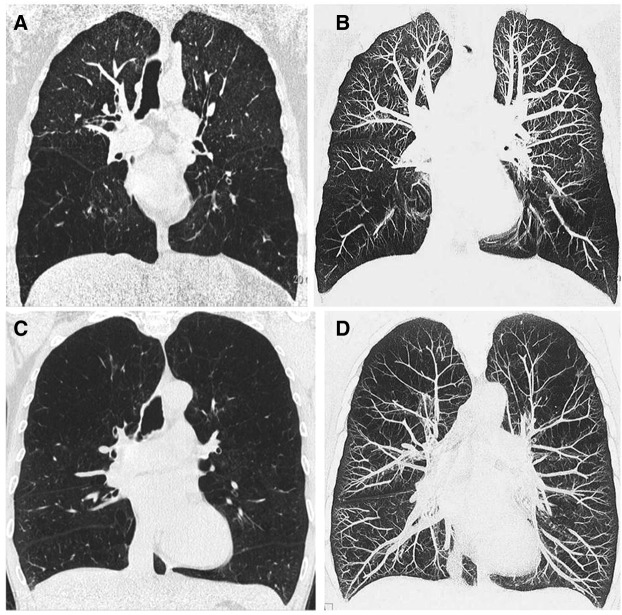
(A and B) Coronal (A) and coronal maximum intensity projection (B) chest computed tomography of a smoker with lower zone–predominant emphysema. Note the distorted and obliterated vasculature in the lower zones. In contrast, the vascular architecture is preserved in the upper zones, which are mostly spared from emphysematous destruction. (C and D) Coronal (C) and coronal maximum intensity projection (D) chest computed tomography of a smoker with severe diffuse emphysema. Note the paucity of distal vessels as well as the elongation and narrowing of the more central pulmonary vasculature. Images courtesy of Dr. Philippe Grenier, Paris, France.
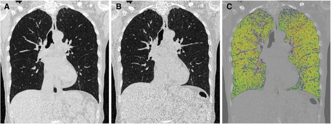
Coronal computed tomography images of a patient with chronic obstructive pulmonary disease in inspiration (A), expiration (B), and parametric response mapping (C). On the parametric response mapping image, green represents normal lung tissue, yellow represents functional small airway disease, and red represents emphysema. This patient has predominantly functional small airway disease involving both lungs.
References
-
- Coxson HO, Leipsic J, Parraga G, Sin DD. Using pulmonary imaging to move chronic obstructive pulmonary disease beyond FEV1. Am J Respir Crit Care Med. 2014;190:135–144. - PubMed
-
- Gould MK, Tang T, Liu IL, Lee J, Zheng C, Danforth KN, Kosco AE, Di Fiore JL, Suh DE. Recent trends in the identification of incidental pulmonary nodules. Am J Respir Crit Care Med. 2015;192:1208–1214. - PubMed
-
- The definition of emphysema: report of a National Heart, Lung, and Blood Institute, Division of Lung Diseases workshop. Am Rev Respir Dis. 1985;132:182–185. - PubMed
-
- Anderson AE, Jr, Hernandez JA, Eckert P, Foraker AG. Emphysema in lung macrosections correlated with smoking habits. Science. 1964;144:1025–1026. - PubMed
Publication types
MeSH terms
Grants and funding
- S10 OD018526/OD/NIH HHS/United States
- K23 HL128936/HL/NHLBI NIH HHS/United States
- R01 HL126838/HL/NHLBI NIH HHS/United States
- HHSN268200900016C/HL/NHLBI NIH HHS/United States
- R01 HL122438/HL/NHLBI NIH HHS/United States
- HHSN268200900013C/HL/NHLBI NIH HHS/United States
- HHSN268200900014C/HL/NHLBI NIH HHS/United States
- K24 HL138188/HL/NHLBI NIH HHS/United States
- T32 HL007749/HL/NHLBI NIH HHS/United States
- R01 HL089897/HL/NHLBI NIH HHS/United States
- HHSN268200900019C/HL/NHLBI NIH HHS/United States
- R01 HL112986/HL/NHLBI NIH HHS/United States
- P30 ES005605/ES/NIEHS NIH HHS/United States
- U01 HL089897/HL/NHLBI NIH HHS/United States
- R01 HL077612/HL/NHLBI NIH HHS/United States
- R01 HL089856/HL/NHLBI NIH HHS/United States
- R35 CA197701/CA/NCI NIH HHS/United States
- U01 HL089856/HL/NHLBI NIH HHS/United States
- HHSN268200900015C/HL/NHLBI NIH HHS/United States
- HHSN268200900018C/HL/NHLBI NIH HHS/United States
- P30 DK054759/DK/NIDDK NIH HHS/United States
- HHSN268200900017C/HL/NHLBI NIH HHS/United States
- HHSN268200900020C/HL/NHLBI NIH HHS/United States
LinkOut - more resources
Full Text Sources
Other Literature Sources
Medical
