

Ultrasound Diagnosis of Calf Injuries
- PMID: 28661826
- PMCID: PMC5496702
- DOI: 10.1177/1941738117696019
Ultrasound Diagnosis of Calf Injuries
Abstract
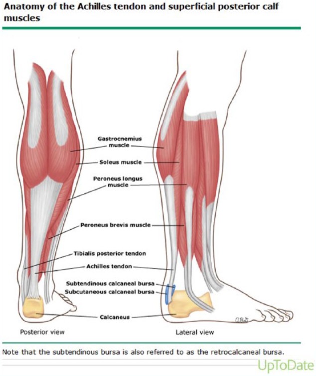
Context: The medial head of the gastrocnemius is the third most commonly strained muscle in elite athletes after the biceps femoris and rectus femoris. The differential diagnosis of posterior calf injury includes musculoskeletal and nonmusculoskeletal causes. Classically, delineation of these injuries from one another relied primarily on historical features and physical examination findings. The utilization of musculoskeletal ultrasound (sonography) has augmented the diagnosis of these injuries by providing dynamic, real-time confirmation.
Evidence acquisition: A review of PubMed, OVID, and MD Consult prior to January 2016 was performed using search terms, including s oleus ultrasound, gastrocnemius ultrasound, and tennis leg. The references of the pertinent articles were further reviewed for other relevant sources.
Study design: Clinical review.
Level of evidence: Level 4.
Results: There have been few reviews to date of calf injuries and the use of sonography in their diagnosis. Prompt diagnosis utilizing ultrasound allows the clinician to focus management on gastrocnemius injury if present. Two-thirds of calf injuries occur at the junction of the fascia between the medial head of the gastrocnemius and the soleus. Injuries to the lateral head of the gastrocnemius occur in up to 14% of patients in some case series, but injury may occur anywhere from the proximal origin to the mid-belly to the fascial junction with the soleus. Numerous injuries to the posterior compartment can mimic gastrocnemius strain, and musculoskeletal ultrasound can aide in their diagnosis by incorporating real-time imaging into the grading of the injury and visual confirmation of physical examination findings.
Conclusion: Acute injury to the posterior compartment of the lower extremity can represent a diagnostic challenge. Medial gastrocnemius strain represents the most common injury of the posterior compartment of the lower extremity. Ultrasound is a useful tool to assist the clinician in determining the specific cause of calf injury, estimate the severity of the injury, and monitor progress of healing. The vast majority of calf injuries can be diagnosed and managed without any additional imaging. Serial diagnostic ultrasound of an injured area allows for direct monitoring of tissue healing and may allow the clinician to more confidently assess response to treatment and help guide return-to-play decisions.
Keywords: calf; gastrocnemius; lower leg; sonography; ultrasound.
Conflict of interest statement
The authors report no potential conflicts of interest in the development and publication of this article. The views expressed in this paper are those of the authors and do not necessarily reflect those of the United States Army or any other section of the Department of Defense.
Figures
References
-
- Al-Himdani S, Talbot C, Kurdy N, Pillai A. Accessory muscles around the foot and ankle presenting as chronic undiagnosed pain. An illustrative case report and review of the literature. Foot (Ednib). 2013;23:154-161. - PubMed
-
- Arner O, Lindholm A. What is tennis leg. Acta Chir Scand. 1958;116:73-75. - PubMed
-
- Athavale SA, Geetha GN, Swathi Morphology of flexor digitorum accessorius muscle. Surg Radiol Anat. 2012;34:367-372. - PubMed
-
- Azar FM. Traumatic disorders. In: Canale ST, Beaty JH, eds. Campbell’s Operative Orthopaedics. Philadelphia, PA: Mosby; 2008:233-234.
-
- Bianchi S, Martolini C, Abdelwahab IF, Derchi LE, Damiani S. Sonographic evaluation of tears of the gastrocnemius medial head (“tennis leg”). J Ultrasound Med. 1998;17:157-162. - PubMed
Publication types
MeSH terms
LinkOut - more resources
Full Text Sources
Other Literature Sources
Medical
Research Materials
