. 2017 Jun 29;12(6):e0179450.
doi: 10.1371/journal.pone.0179450. eCollection 2017.

Activation of complement factor B contributes to murine and human myocardial ischemia/reperfusion injury

Affiliations

Activation of complement factor B contributes to murine and human myocardial ischemia/reperfusion injury

Nicholas Chun et al. PLoS One. .

Abstract

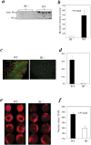
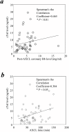
The pathophysiology of myocardial injury that results from cardiac ischemia and reperfusion (I/R) is incompletely understood. Experimental evidence from murine models indicates that innate immune mechanisms including complement activation via the classical and lectin pathways are crucial. Whether factor B (fB), a component of the alternative complement pathway required for amplification of complement cascade activation, participates in the pathophysiology of myocardial I/R injury has not been addressed. We induced regional myocardial I/R injury by transient coronary ligation in WT C57BL/6 mice, a manipulation that resulted in marked myocardial necrosis associated with activation of fB protein and myocardial deposition of C3 activation products. In contrast, in fB-/- mice, the same procedure resulted in significantly reduced myocardial necrosis (% ventricular tissue necrotic; fB-/- mice, 20 ± 4%; WT mice, 45 ± 3%; P < 0.05) and diminished deposition of C3 activation products in the myocardial tissue (fB-/- mice, 0 ± 0%; WT mice, 31 ± 6%; P<0.05). Reconstitution of fB-/- mice with WT serum followed by cardiac I/R restored the myocardial necrosis and activated C3 deposition in the myocardium. In translational human studies we measured levels of activated fB (Bb) in intracoronary blood samples obtained during cardio-pulmonary bypass surgery before and after aortic cross clamping (AXCL), during which global heart ischemia was induced. Intracoronary Bb increased immediately after AXCL, and the levels were directly correlated with peripheral blood levels of cardiac troponin I, an established biomarker of myocardial necrosis (Spearman coefficient = 0.465, P < 0.01). Taken together, our results support the conclusion that circulating fB is a crucial pathophysiological amplifier of I/R-induced, complement-dependent myocardial necrosis and identify fB as a potential therapeutic target for prevention of human myocardial I/R injury.


Conflict of interest statement

Competing Interests: The authors have declared that no competing interests exist.

Figures

Fig 1
Fig 1. Factor B knockout mice experienced reduced myocardial necrosis and complement C3 deposition.
fB-/- mice and WT were used in a myocardial I/R model. The left anterior descending (LAD) coronary artery was occluded for 1 hr then reperfused for 24 hrs. Propidium iodide and blue fluorescent microspheres (BFM) (the latter after re-occlusion of the LAD) were injected in vivo just prior to heart harvesting to delineate the necrotic tissue and the tissue lacking circulation and therefore at risk for necrosis, respectively. (a) Circulating fB in the blood was significantly activated in WT mice (n = 4) but not in fB-/- mice (n = 4). Serum obtained from cardiac puncture at the end of reperfusion was analyzed by Western blotting as described in Materials and Methods, Section 3. Each lane in the blot represented a separate mouse. Arrows indicate fB and Bb fragments. (b) The bar chart summarizes the relative intensities of Bb fragments. Bars represent Means ± SEM (* indicates P<0.05 compared with WT controls). (c) Cryosections prepared from the heart slices were stained with a FITC-tagged anti-C3 antibody. (d) Bar graph: bars indicate the percentage of total area that is C3 positive. (e) Necrotic tissue (bright red fluorescence) was visualized under a fluorescent microscope immediately after the harvest of hearts, using a 2x objective lens, in slices obtained by dividing the heart into four (top and bottom of each slice are adjacent in the figure). Non-ischemic tissue was defined by the blue fluorescence of BFM, the non-fluorescing tissue constituting weight of tissue at risk (WAR) (n = 7 per group). (f) Bar graph: Necrotic area expressed as % WAR as defined in the Materials and Methods, Section 2.
Fig 2
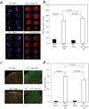
Fig 2. Factor B from circulation contributed myocardial necrosis in cardiac I/R.
fB-/- mice (n = 4) were re-constituted i.v. with 500ul WT serum (first 250ul i.v. 1 hour prior to surgery; second 250 μl i.v. 40 minutes prior to surgery) to provide an extracellular source of fB. WT mice (n = 4; similarly injected) were used as positive controls. Animals were subjected to myocardial IR (1 h ischemia/24 h reperfusion). Sham operated fB-/- (n = 5) or WT mice (n = 5) were included as negative controls. (a) Myocardial necrosis was determined as done in Fig 1. (b) Bar graph: Necrotic area expressed as % WAR as defined in Fig 1. (c) Heart cryosections were stained with a FITC-tagged anti-C3 antibody. (d) Bar graph: bars indicate the percentage of total area that is C3 positive.
Fig 3
Fig 3. mRNA expression of fB in the WT hearts after IR.
RNA were isolated from WT hearts without surgery (naïve group; n = 3), or sham operation (n = 10), or I/R operation (n = 12). cDNA were synthesized, reverse transcribed, and real-time RT-PCR were performed as described in Method Section.
Fig 4
Fig 4. Activation of fB in the coronary circulation after AXCL.
(a) Bb levels increased in the coronary circulation after the AXCL of human cardiac surgery. Coronary sinus blood was collected prior to AXCL and 5 minutes after AXCL termination. Bb levels were determined by ELISA using an antibody which detects the Bb component of activated fB but not native fB, nor the other fragment of fB activation, Ba. Statistical significances were analyzed as described in Materials and Methods, Section 8. Box-charts were plotted using SigmaPlot software. The boundary of the box closest to zero indicates the 25th percentile, while the boundary of the box farthest from zero indicates the 75th percentile. Error bars above and below the box indicate the 90th and 10th percentiles. The two filled circles above and below the box indicate the 95th and 5th percentiles. The solid line within the box marks the median, and the dotted line marks the mean (average). N = 56; * indicates statistical significance (P < 0.05). (b) Plasma from the coronary sinus blood obtained before the start of AXCL and 5 minutes after AXCL cessation was analyzed by Western blotting using a polyclonal antibody that detects fB and Bb. A representative blot is depicted showing fB positive bands from a patient’s plasma. Arrows indicate fB and Bb fragments. Band intensities on scanned images of such blots were quantified and normalized to the 93 kDa fB band of a control plasma and expressed as relative intensity. (c) A bar chart summarizes the relative intensities of Bb fragments.
Fig 5
Fig 5
(a) The increases in coronary blood Bb levels immediately after AXCL correlated significantly with postoperative increases in cTn1 levels. (b) The increases in postoperative increases in cTn1 levels correlated significantly with AXCL time. Univariate analyses were carried out as described in Method. * indicates statistical significance.

References

    1. Eltzschig HK, Eckle T. Ischemia and reperfusion—from mechanism to translation. Nature medicine. 2011;17(11):1391–401. Epub 2011/11/09. doi: 10.1038/nm.2507 ;. - DOI - PMC - PubMed
    1. Millington TM, Madsen JC. Innate immunity and cardiac allograft rejection. Kidney Int Suppl. 2010;(119):S18–21. Epub 2010/12/01. doi: 10.1038/ki.2010.417 ;. - DOI - PMC - PubMed
    1. Lee JC, Christie JD. Primary graft dysfunction. Clin Chest Med. 2011;32(2):279–93. Epub 2011/04/23. doi: 10.1016/j.ccm.2011.02.007 . - DOI - PubMed
    1. Schmauss D, Weis M. Cardiac allograft vasculopathy: recent developments. Circulation. 2008;117(16):2131–41. Epub 2008/04/23. doi: 10.1161/CIRCULATIONAHA.107.711911 . - DOI - PubMed
    1. Arcasoy SM, Fisher A, Hachem RR, Scavuzzo M, Ware LB. Report of the ISHLT Working Group on Primary Lung Graft Dysfunction part V: predictors and outcomes. The Journal of heart and lung transplantation: the official publication of the International Society for Heart Transplantation. 2005;24(10):1483–8. Epub 2005/10/08. doi: 10.1016/j.healun.2004.11.314 . - DOI - PubMed

Substances