Review
. 2017 Jun 29;12(1):50.
doi: 10.1186/s13024-017-0192-x.

Glial contributions to neurodegeneration in tauopathies

Affiliations
Review

Glial contributions to neurodegeneration in tauopathies

Cheryl E G Leyns et al. Mol Neurodegener. .

Abstract

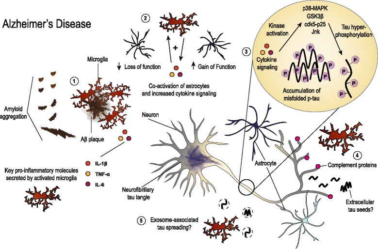
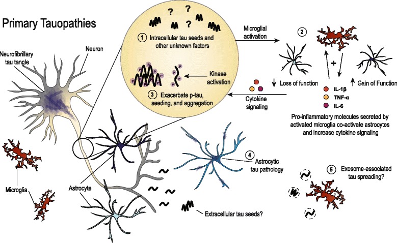
Tauopathies are a broad set of neurodegenerative dementias characterized by aggregation of the tau protein into filamentous inclusions that can be found in neurons and glial cells. Activated microglia, astrocytes and elevated levels of proinflammatory molecules are also pathological hallmarks that are found in brain regions affected by tau pathology. There has been abundant research in recent years to understand the role of gliosis and neuroinflammation in neurodegenerative diseases, particularly in Alzheimer's disease (AD) which is the most common form of dementia. AD is a tauopathy characterized by both extracellular amyloid-β plaques in addition to intracellular neurofibrillary tangles and neuropil threads containing aggregated tau protein. Accumulating evidence suggests that neuroinflammation offers a possible mechanistic link between these pathologies. Additionally, there appears to be a role for neuroinflammation in aggravating tau pathology and neurodegeneration in tauopathies featuring tau deposits as the predominant pathological signature. In this review, we survey the literature regarding inflammatory mechanisms that may impact neurodegeneration in AD and related tauopathies. We consider a physical role for microglia in the spread of tau pathology as well as the non-cell autonomous effects of secreted proinflammatory cytokines, specifically interleukin 1 beta, interleukin 6, tumor necrosis factor alpha and complement proteins. These molecules appear to have direct effects on tau pathophysiology and overall neuronal health. They also indirectly impact neuronal homeostasis by altering glial function. We conclude by proposing a complex role for gliosis and neuroinflammation in accelerating the progression of AD and other tauopathies.

Keywords: Alzheimer’s disease; Astrocyte; Gliosis; Microglia; Neurodegeneration; Neuroinflammation; Tau; Tauopathy.


Conflict of interest statement

Ethics approval and consent to participate

Not applicable.

Consent for publication

Not applicable.

Competing interests

CEGL and DMH are listed as inventors on a patent licensed by Washington University to C2N Diagnostics on the therapeutic use of anti-tau antibodies. DMH co-founded and is on the scientific advisory board of C2N Diagnostics, LLC. C2N Diagnostics, LLC has licensed certain anti-tau antibodies to AbbVie for therapeutic development. DMH is on the scientific advisory board of Proclara and consults for Genentech, Eli Lilly, Denali, AbbVie, and GlaxoSmithKline. The authors have no additional financial interests.

Publisher’s Note

Springer Nature remains neutral with regard to jurisdictional claims in published maps and institutional affiliations.

Figures

Fig. 1
Fig. 1
Illustration summarizing the hypothesized roles of gliosis and neuroinflammation in AD. Aggregation of Aβ likely stimulates microglia early in disease and may instigate initial neuroinflammation (1). While gliosis has been shown to be beneficial in reducing plaque burden and mitigating amyloid-associated pathologies, long-term stimulation creates a permissive environment for chronic neuroinflammation. Pro-inflammatory cytokines such as IL-1β, TNF-α and IL-6 further activate microglia and astrocytes, leading to both loss and gain of functions (2). These molecules have been shown to disrupt neuronal homeostasis and alter tau biology. Cytokine signaling has been linked to activation of kinases that phosphorylate tau, which may incite early tau dysfunction and ultimately influence misfolding and accumulation (3). Concurrently, there is deposition of complement proteins at neuronal synapses that can signal microglial pruning and initiate synapse loss (4). Microglia have also been implicated in facilitating the spread of tau via exosomes (5). Additionally, neuronal activity-dependent release of extracellular, misfolded tau may incite neuronal dysfunction or spread of tau pathology along synaptically connected neuronal populations. Ultimately, gliosis and chronic neuroinflammation combine with plaque and tangle pathologies to drive neurodegeneration in AD
Fig. 2
Fig. 2
Depiction of the roles that have been described for glial cells in primary tauopathies. In the absence of amyloid pathology, early microgliosis may be initiated by neuronal tau seeds (1), though this remains to be thoroughly tested. Tau seeding has been found early in mouse models of tauopathy, and secreted, extracellular tau also possibly has seeding capability. Either direct or indirect consequences from tau seeds may be responsible for instigating the early microgliosis reported in tau mouse models. Activated microglia then secrete pro-inflammatory cytokines which further exacerbates microgliosis and co-activates astrocytes leading to toxic loss and gain of functions (2). Similar to the role proposed for AD, gliosis and inflammatory signaling can impact tau phosphorylation and possibly enhance misfolding and aggregation (3). In addition, astrocytic tau pathology characterizes several primary tauopathies such as PSP and CBD (4), though the functional consequences of the different aggregate phenotypes that are observed remain unknown. Spread of toxic tau species via microglial-associated exosomes is also a possible mechanism in primary tauopathies (5). Together, chronic neuroinflammation combined with tau pathology diminishes neuronal health and worsens neurodegeneration

References

    1. Spillantini MG, Goedert M. Tau pathology and neurodegeneration. Lancet Neurol. 2013;12:609–622. doi: 10.1016/S1474-4422(13)70090-5. - DOI - PubMed
    1. Arendt T, Stieler JT, Holzer M. Tau and tauopathies. Brain Res Bull. 2016;126:238–292. doi: 10.1016/j.brainresbull.2016.08.018. - DOI - PubMed
    1. Musiek ES, Holtzman DM. Three dimensions of the amyloid hypothesis: time, space and “wingmen.”. Nat Neurosci. 2015;18:800–806. doi: 10.1038/nn.4018. - DOI - PMC - PubMed
    1. Wyss-Coray T, Mucke L. Inflammation in neurodegenerative disease--a double-edged sword. Neuron. 2002;35:419–432. doi: 10.1016/S0896-6273(02)00794-8. - DOI - PubMed
    1. Ransohoff RM. How neuroinflammation contributes to neurodegeneration. Science. 2016;353:777–783. doi: 10.1126/science.aag2590. - DOI - PubMed