Review
. 2017 Aug 2;25(8):1757-1768.
doi: 10.1016/j.ymthe.2017.06.003. Epub 2017 Jun 26.

Quantitative Imaging Approaches to Study the CAR Immunological Synapse

Affiliations
Review

Quantitative Imaging Approaches to Study the CAR Immunological Synapse

Malini Mukherjee et al. Mol Ther. .

Abstract

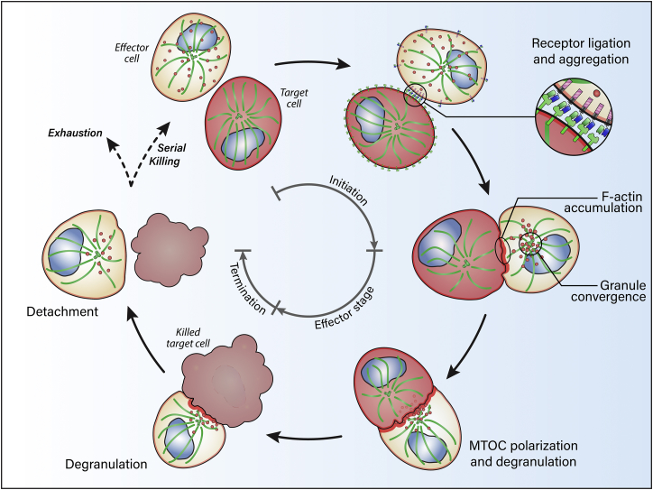
The lytic immunological synapse (IS) is a discrete structural entity formed after the ligation of specific activating receptors that leads to the destruction of a cancerous cell. The formation of an effector cell IS in cytotoxic T lymphocytes or natural killer cells is a hierarchical and stepwise rearrangement of structural and signaling components and targeted release of the contents of lytic granules. While recent advances in the generation and testing of cytotoxic lymphocytes expressing chimeric antigen receptors (CARs) has demonstrated their efficacy in the targeted lysis of tumor targets, the contribution and dynamics of IS components have not yet been extensively investigated in the context of engineered CAR cells. Understanding the biology of the CAR IS will be a powerful approach to efficiently guide the engineering of new CARs and help identify mechanistic problems in existing CARs. Here, we review the formation of the lytic IS and describe quantitative imaging-based measurements using multiple microscopy techniques at a single cell level that can be used in conjunction with established population-based assays to provide insight into the important cytotoxic function of CAR cells. The inclusion of this approach in the pipeline of CAR product design could be a novel and valuable innovation for the field.

Keywords: CAR; chimeric antigen receptor; confocal microscopy; immune synapse; live imaging microscopy; quantitative image analysis.


Figures

None
Graphical abstract
Figure 1
Figure 1
Evolution of CAR Designs Schematic model of typical CAR designs each representing a different generation (main panel). The elementary blocks used for their design are put back into their original context in the insets: the top panel shows the domains of a monoclonal antibody used to create the target-specific scFv domain of the CAR, while the bottom inset depicts the signaling receptors (CD3ζ, CD28, 4-1BB, OX40) that provide the CAR intracellular signaling domain. The first-generation CAR has a single activation signal, while the second-generation CAR integrates activation and a single co-stimulatory signal in cis. The third generation of CARs includes more than one co-stimulatory signal that can be presented in cis or in trans as a co-stimulatory receptor (CCR) when paired with a first-generation CAR. mAb, monoclonal antibody.
Figure 2
Figure 2
Schematic Representation of the Critical Steps of the Killing of a Sensitive Target by a CAR-Expressing Effector Cell The “initiation” stage includes the recognition and the ligation of the CAR receptor with its ligand at the surface of the target cell. Following intracellular signaling, the receptors at the surface of the effector converge and aggregate toward the interface between the two cells where the IS is forming. The following stage is referred to as the “effector stage” and encompasses multiple discrete steps: the accumulation of filamentous actin at the IS, the convergence of the lytic granules around the microtubule organizing center (MTOC), the polarization of the granule-loaded MTOC against the plasma membrane at the IS and, ultimately, the release of the cytolytic granules in the IS cleft leading to the killing of the target cell. The final stage closing the cycle is the “termination” stage: the target cell carries on its apoptotic process and then separates from the effector, allowing the latter to proceed with the killing of another target cell, a mechanism called serial killing. If the effector cannot regenerate its granule content, it remains depleted and enters a stage of exhaustion. Note that the receptor and ligands at the cell surface of the cells are only depicted in one panel for clarity purposes but are present at all stages.
Figure 3
Figure 3
Typical Workflow of CAR Design and Proposed Imaging-Based Interrogations The middle pipeline describes the successive steps leading to the production of an efficient CAR that can be evaluated in clinical trials. On the left side are the most common bulk assays currently in use to optimize and test CAR products. On the right side of the central stem is the list of microscopy-based experiments that we propose to gain a better insight into the mechanism of action of the current CAR. The quantitative results of these assays can be used iteratively to optimize the various stages of the CAR design pipeline.
Figure 4
Figure 4
Simplified Schematics alongside Representative Volume Projections of Microscopy Data of CAR NK Cell/Target Cell Conjugates Demonstrating Four Quantifiable Parameters of IS Formation (A) CAR-antigen aggregation, (B) F-actin accumulation, (C) granule convergence, and (D) MTOC polarization. (A) The first panel series describes the accumulation of CAR at the IS formed with the target cell as one of the first measurable steps in CAR-mediated cytotoxicity. The representative fluorescence microscopy image shows the region overlapping between the CD19 antigen of the target cell (blue) and the mEmeraldGFP-CD19-CAR of the CAR T cell (green). A quantifiable parameter can be extracted by measuring the fluorescence intensity of the CD19-CAR at the IS versus the total fluorescence intensity in the effector and expressed as a ratio. (B) The second panel series shows accumulation of F-actin at the IS (green) as the second measurable step in CAR-mediated cytotoxicity. In the microscopy image, a region of interest (ROI) is drawn to indicate the accumulation of filamentous actin at the interface between effector and target cells. Once again, this can be quantified with a ratio between the fluorescence intensity in this region compared to the total intensity in the entire effector cell. (C) The third panel series illustrates the convergence of the lytic granules around the MTOC. The distance from each individual granule (here stained for perforin, red) to the MTOC (here stained for pericentrin, blue) is materialized by a red line in the schematic and the example dataset. The mean distance between all the granules and the MTOC in each cell is an accurate descriptor of their state of convergence. (D) The bottom panel series shows polarization of the MTOC to the IS, which marks the fourth readily measurable stage. The length of the shortest line drawn from the MTOC (here stained for pericentrin, blue) to the IS (here materialized by a staining for filamentous actin, green) measures the progression of polarization. The example dataset in the rightmost panel shows a MTOC together with converged granules approaching the surface of the IS. Scale bars represent 1 μm.

References

    1. Gross G., Gorochov G., Waks T., Eshhar Z. Generation of effector T cells expressing chimeric T cell receptor with antibody type-specificity. Transplant. Proc. 1989;21:127–130. - PubMed
    1. Irving B.A., Weiss A. The cytoplasmic domain of the T cell receptor zeta chain is sufficient to couple to receptor-associated signal transduction pathways. Cell. 1991;64:891–901. - PubMed
    1. Eshhar Z., Waks T., Gross G., Schindler D.G. Specific activation and targeting of cytotoxic lymphocytes through chimeric single chains consisting of antibody-binding domains and the gamma or zeta subunits of the immunoglobulin and T-cell receptors. Proc. Natl. Acad. Sci. USA. 1993;90:720–724. - PMC - PubMed
    1. Imai C., Mihara K., Andreansky M., Nicholson I.C., Pui C.H., Geiger T.L., Campana D. Chimeric receptors with 4-1BB signaling capacity provoke potent cytotoxicity against acute lymphoblastic leukemia. Leukemia. 2004;18:676–684. - PubMed
    1. Fedorov V.D., Sadelain M., Kloss C.C. Novel approaches to enhance the specificity and safety of engineered T cells. Cancer J. 2014;20:160–165. - PMC - PubMed

MeSH terms