. 2017 Sep 1;429(1):213-224.
doi: 10.1016/j.ydbio.2017.06.025. Epub 2017 Jun 27.

Wbp2nl has a developmental role in establishing neural and non-neural ectodermal fates

Affiliations

Wbp2nl has a developmental role in establishing neural and non-neural ectodermal fates

Alexander Marchak et al. Dev Biol. .

Abstract

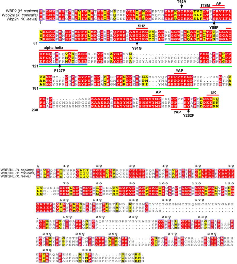
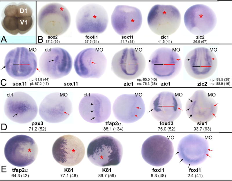
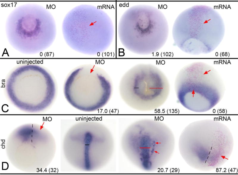
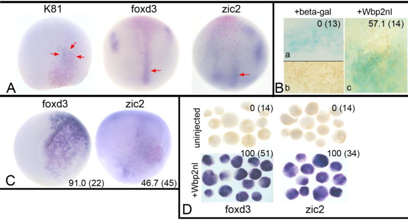
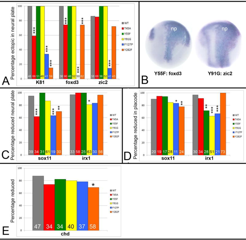
In many animals, maternally synthesized mRNAs are critical for primary germ layer formation. In Xenopus, several maternal mRNAs are enriched in the animal blastomere progenitors of the embryonic ectoderm. We previously identified one of these, WW-domain binding protein 2 N-terminal like (wbp2nl), that others previously characterized as a sperm protein (PAWP) that promotes meiotic resumption. Herein we demonstrate that it has an additional developmental role in regionalizing the embryonic ectoderm. Knock-down of Wbp2nl in the dorsal ectoderm reduced cranial placode and neural crest gene expression domains and expanded neural plate domains; knock-down in ventral ectoderm reduced epidermal gene expression. Conversely, increasing levels of Wbp2nl in the neural plate induced ectopic epidermal and neural crest gene expression and repressed many neural plate and cranial placode genes. The effects in the neural plate appear to be mediated, at least in part, by down-regulating chd, a BMP antagonist. Because the cellular function of Wbp2nl is not known, we mutated several predicted motifs. Expressing mutated proteins in embryos showed that a putative phosphorylation site at Thr45 and an α-helix in the PH-G domain are required to ectopically induce epidermal and neural crest genes in the neural plate. An intact YAP-binding motif also is required for ectopic epidermal gene expression as well as for down-regulating chd. This work reveals novel developmental roles for a cytoplasmic protein that promotes epidermal and neural crest formation at the expense of neural ectoderm.

Keywords: Neural crest; Neural plate; PAWP; Placodes; foxd3; zic2.


Figures

Figure 1
Figure 1. Amino acid sequence alignments of Xenopus Wbp2nl to human WBP2 and WBP2NL
(A) Xenopus Wbp2nl (NP_001088037.1 [laevis] and NP_001107397.1 [tropicalis]) is highly similar to human WBP2 (NP_036610.2). Blue bar indicates the predicted PH-G domain and green bar indicates the predicted WWbp domain. Highly conserved motifs and α-helix are indicated by red bars. Five single amino acid mutations that were experimentally tested for function are indicated by black arrows (T45A, Y55F, Y91G, F127P, Y282F). AP, adaptor protein complex; ER, endoplasmic reticulum retention signal; ITSM, immunoreceptor tyrosine-based switch motif; SH2, Scr Homology domain 2; YAP, Yes associated protein binding motif. (B) Comparison of human WBP2NL (AAH22546.1) to Xenopus sequences shows large regions of disimilarity. Another human WBP2NL entry (Q6ICG8.1) is identical to AAH22546.1 except there is a glutamine at position 285 instead of a histidine. (C) Four examples of cells immunostained for Myc-Wbp2nl (green). Nuclei are stained with DAPI (blue). Note that staining is perinuclear (red arrows, and in cytoplasmic tubular structures (blue arrows) consistent with affiliation with Endoplasmic reticulum. Red asterisks denote intracellular yolk platelets.
Figure 2
Figure 2. Reduction of Wbp2nl affects ectodermal tissues
(A) An 8-cell Xenopus laevis embryo. The dorsal-animal blastomere (D1) gives rise to the neural plate and the ventral-animal blastomere (V1) gives rise to the epidermis. Border zone derivatives (neural crest and cranial placodes) descend from the lateral regions of both blastomeres (Moody and Kline, 1990). (B) Knock-down of Wbp2nl causes a decrease in the expression (loss of blue reaction product) of neural ectodermal genes on the MO-injected side (asterisk) of gastrula stage embryos. sox2 is a vegetal view with dorsal to the top; the rest are dorsal views with animal to the top. (C) By neural plate stages, neural gene domains are expanded; compare the width of the neural plate on MO-injected side (red bar) to control side (black bar). (See also D). The PPE domains of sox11 and the neural crest domains of zic1 and zic2 are reduced on MO-injected sides (red arrows) compared to control sides (black arrows). Anterior views with dorsal to the top. (D) At neural plate stages, neural crest markers (pax3, tfap2α, foxd3) and an early PPE marker (six1) are reduced on the MO-injected side (red arrows) compared to control sides (black arrows). Also note wider neural plates (red bars). pax3 and tfap2α are anterior-side views; foxd3 and six1 are anterior views. (E) Two epidermal genes are reduced at the sites of Wbp2nl knock-down (asterisks) at gastrula (tfap2α, K81 right image; both are animal pole views) and neural plate (K81 left image; side view with dorsal to top and anterior to right) stages. In contrast, the animal pole expression of foxi1 in the gastrula (left image) is not altered. Rarely, the PPE expression of foxi1 (right image; anterior view with dorsal to top) is reduced (red arrows) after Wbp2nl knockdown. Frequencies of the phenotypes and sample sizes (n) are given in each panel.
Figure 3
Figure 3. Increasing Wbp2nl levels alters ectodermal gene expression
(A) Increased Wbp2nl causes ectopic expression of epidermal keratin (K81) in the neural ectoderm (ne) at gastrula stages (left) but not at neural plate (np) stages (right). Pink nuclei identify the cells that are expressing excess Wbp2nl and the black arrow indicated the posterior extent of the clone. A–D, dorsal views with animal to top. (B) Increased Wbp2nl does not induce ectopic foxi1 in the ne. (C) Increased Wbp2nl causes ectopic expression of foxd3 in the neural plate (np). (D) Increased Wbp2nl causes ectopic expression of zic2 in the neural plate. (E–G) Increased Wbp2nl does not cause ectopic neural plate expression of pax3, zic1 or six1 (dorsal views with animal to top). In fact, zic1 neural plate expression is reduced on the injected side (F, right image, anterior view, dorsal to top). (H) Increased Wbp2nl does not alter foxd4l1 neural ectoderm expression. Vegetal-dorsal view. (I) Neural ectoderm expression of sox2 in the gastrula is reduced compared to lateral regions (black arrows) that do not contain Wbp2nl-expressing cells (pink nuclei). Dorsal view, animal to top. (J) Both the neural plate (red bar) and placode (red arrows) domains of irx1 are reduced by increased Wbp2nl compared to control side (black bar). Dorsal view, anterior to top. (K) Both the neural plate and placode (red arrows) expression of sox11 are reduced by increased Wbp2nl. Anterior view, dorsal to top. (L) The placode (red arrows) domain of six1 is reduced by increased Wbp2nl, compared to control uninjected side (black arrows). Side views, dorsal to top. (M) The placode (red arrows) domain of sox11 is reduced by increased Wbp2nl compared to control uninjected side (black arrows). Note reduced neural plate (np) staining as well. Side views, dorsal to top. Frequencies of the phenotypes and sample sizes (n) are given in each panel.
Figure 4
Figure 4. Altered Wbp2nl levels affect mesoderm but not endoderm
(A) At gastrula stages, sox17 expression surrounding the ventral blastopore on the MO-injected side of an embryo (left image, vegetal view) is not discernably different from control side. Increased Wbp2nl in the animal pole ectoderm (right image, animal view; clone marked by pink nuclei and red arrow) does not ectopically induce sox17. (B) At gastrula stages, edd expression surrounding the ventral blastopore on the MO-injected side (left image, vegetal view) is not discernably different from control side. Increased Wbp2nl in the neural ectoderm (right image, dorsal view; clone marked by pink nuclei and red arrow) does not ectopically induce edd. (C) At gastrula stages, mesoderm expression of bra encircles the blastopore in uninjected control embryos (left image, vegetal view); knockdown of Wbp2nl causes a loss of bra expression (left middle image, vegetal view, red arrow). In contrast, at neural plate stages (right middle image, vegetal view), we detect a posterior expansion (red bar) of bra expression on the knock-down side. At gastrula stages, increased Wbp2nl (right image, dorsal view; clone marked by pink nuclei between red arrows) does not alter endogenous bra expression nor ectopically induce it. (D) At gastrula stages, mesoderm expression of chd is reduced by Wbp2nl knock-down (red arrow, left image, vegetal view; dashed line indicates dorsal midline). In contrast, at neural plate stages, chd is expanded (compare uninjected control pattern [left middle image] to MO-injected pattern [right middle image]; dorsal views with anterior to the top). Red bar shows a wider domain and red arrows denote ectopic expression. At gastrula stages, increased Wbp2nl (right image, dorsal view, anterior to top; dashed line indicates dorsal midline) also reduces endogenous chd expression (red arrow). Frequencies of the phenotypes and sample sizes (n) are given in each panel.
Figure 5
Figure 5. BMP signaling underlies Wbp2nl-induced ectopic expression phenotypes
(A) The ectopic expression of K81, foxd3 and zic2 are minimized by co-expression of Chd, a BMP antagonist. Red arrows point to weak posterior ectopic gene expression observed in those few embryos that were positively scored. (B) Immunostaining for phosphorylated SMAD 1/5/8, an indicator of BMP signaling when located in the nucleus. Only embryos in which ventral cells, which are subject to high levels of endogenous BMP signaling, showed nuclear staining were analyzed (b). No embryo injected only with lineage tracer (blue) showed nuclear staining in the neural plate (a), whereas a majority of embryos injected with lineage tracer plus wbp2nl mRNA showed nuclear staining (c). (C) Expression of Wbp2nl in the ventral epidermis causes ectopic expression of two neural crest genes (foxd3, zic2). Pink nuclei (lineage tracer) indicate the Wbp2nl containing cells. Ventral views, anterior to the top. (D) Ventral animal blastomeres were dissected from the 16-cell stage embryo and cultured in simple salt medium. Those dissected from uninjected control embryos never expressed foxd3 or zic2, whereas those that express Wbp2nl (red nuclei) always expressed these genes. Frequencies of the phenotypes and sample sizes (n) are given in each panel.
Figure 6
Figure 6. Mutations in predicted functional domains of Wbp2nl, indicated in Figure 1A, affect its ability to change embryonic gene expression
(A) Percentages of embryos that show ectopic neural plate expression of indicated genes after injection of wild type (WT) or mutated mRNAs: T45A, Y55F and Y91G disrupt putative phosphorylation sites in the PH-G domain; F127P disrupts an a-helix in the PH-G domain; Y282F disrupts a putative YAP binding site at the C-terminus (see Figure 1A). Sample sizes are numbers at the base of each bar. *, p<0.05; **, p<0.01; ***, p<0.005, Chi-squared statistic. (B) Two examples of more intense and broader ectopic expression domains of foxd3 or zic2 in the neural plate (np) after expression of two mutant proteins (Y55F; Y91G). Compare to WT images (Fig. 4C, D). Dorsal views, anterior to the top. (C) Percentages of embryos that show reduced neural plate expression of indicated genes after expression of wild type (WT) or mutated proteins. (D) Percentages of embryos that show reduced placode expression of indicated genes after expression of wild type (WT) or mutated proteins. (E) Percentages of embryos that show reduced chd expression after expression of wild type (WT) or mutated proteins.

Similar articles

Cited by

References

    1. Aarabi M, Qin Z, Xu W, Mewburn J, Oko R. Sperm-borne protein, PAWP, initiates zygotic development in Xenopus laevis by eliciting intracellular calcium release. Mol. Repro Dev. 2010;77:249–256. - PubMed
    1. Asaoka Y, Hata S, Namae M, Furutani-Seiki M, Nishina H. The Hippo pathway controls a switch between retinal progenitor cell proliferation and photoreceptor cell differentiation in zebrafish. PLoS One. 2014;9:e97365. - PMC - PubMed
    1. Begley MJ, Taylor GS, Kim SA, Veine DM, Dixon JE, Stuckey JA. Crystal structure of a phosphoinositide phosphatase, MTMR2: insights into myotubular myopathy and Charcot-Marie-Tooth syndrome. Mol Cell. 2003;12:1391–402. - PubMed
    1. Blum M, De Robertis EM, Wallingford JB, Niehrs C. Morpholinos: Antisense and sensibility. Dev. Cell. 2015;35:145–149. - PubMed
    1. Buchan DWA, Minneci F, Nugent TCO, Bryson K, Jones DT. Scalable web services for the PSIPRED Protein Analysis Workbench. Nucleic Acids Res. 2013;41(W1):W340–W348. - PMC - PubMed

MeSH terms

LinkOut - more resources