

Behavioral Consequences of a Bifacial Map in the Mouse Somatosensory Cortex
- PMID: 28663199
- PMCID: PMC5546400
- DOI: 10.1523/JNEUROSCI.0598-17.2017
Behavioral Consequences of a Bifacial Map in the Mouse Somatosensory Cortex
Abstract
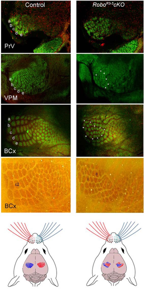
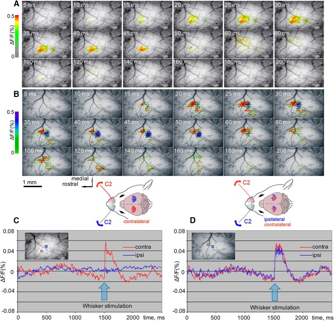
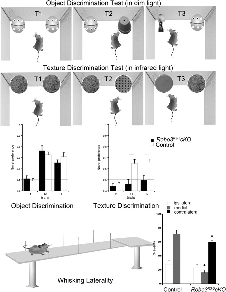
The whisker system is an important sensory organ with extensive neural representations in the brain of the mouse. Patterned neural modules (barrelettes) in the ipsilateral principal sensory nucleus of the trigeminal nerve (PrV) correspond to the whiskers. Axons of the PrV barrelette neurons cross the midline and confer the whisker-related patterning to the contralateral ventroposteromedial nucleus of the thalamus, and subsequently to the cortex. In this way, specific neural modules called barreloids and barrels in the contralateral thalamus and cortex represent each whisker. Partial midline crossing of the PrV axons, in a conditional Robo3 mutant (Robo3R3-5cKO) mouse line, leads to the formation of bilateral whisker maps in the ventroposteromedial, as well as the barrel cortex. We used voltage-sensitive dye optical imaging and somatosensory and motor behavioral tests to characterize the consequences of bifacial maps in the thalamocortical system. Voltage-sensitive dye optical imaging verified functional, bilateral whisker representation in the barrel cortex and activation of distinct cortical loci following ipsilateral and contralateral stimulation of the specific whiskers. The mutant animals were comparable with the control animals in sensorimotor tests. However, they showed noticeable deficits in all of the whisker-dependent or -related tests, including Y-maze exploration, horizontal surface approach, bridge crossing, gap crossing, texture discrimination, floating in water, and whisking laterality. Our results indicate that bifacial maps along the thalamocortical system do not offer a functional advantage. Instead, they lead to impairments, possibly due to the smaller size of the whisker-related modules and interference between the ipsilateral and contralateral whisker representations in the same thalamus and cortex.SIGNIFICANCE STATEMENT The whisker sensory system plays a quintessentially important role in exploratory behavior of mice and other nocturnal rodents. Here, we studied a novel mutant mouse line, in which the projections from the brainstem to the thalamus are disrupted. This led to formation of bilateral whisker maps in both the thalamus and the cortex. The two whisker maps crowd in a space normally devoted to the contralateral map alone and in a nonoverlapping fashion. Stimulation of the whiskers on either side activates the corresponding region of the map. Mice with bilateral whisker maps perform well in general sensorimotor tasks but show poor performance in specific tests that require whisker-dependent tactile discrimination. These observations indicate that contralateral, instead of bilateral, representation of the sensory space plays a critical role in acuity and fine discrimination during somesthesis.
Keywords: Robo3; barrel cortex; barreloids; midline crossing defects; optical imaging; whisker-sensitive behaviors.
Copyright © 2017 the authors 0270-6474/17/377209-10$15.00/0.
Conflict of interest statement
The authors declare no competing financial interests.
Figures
Similar articles
-
Bilateral Whisker Representations in the Primary Somatosensory Cortex in Robo3cKO Mice Are Reflected in the Primary Motor Cortex.Neuroscience. 2024 Apr 19;544:128-137. doi: 10.1016/j.neuroscience.2024.02.031. Epub 2024 Mar 5. Neuroscience. 2024. PMID: 38447690 Free PMC article.
-
Layers 3 and 4 Neurons of the Bilateral Whisker-Barrel Cortex.Neuroscience. 2022 Jul 1;494:140-151. doi: 10.1016/j.neuroscience.2022.05.018. Epub 2022 May 20. Neuroscience. 2022. PMID: 35598701 Free PMC article.
-
Thalamic NMDA receptor function is necessary for patterning of the thalamocortical somatosensory map and for sensorimotor behaviors.J Neurosci. 2014 Sep 3;34(36):12001-14. doi: 10.1523/JNEUROSCI.1663-14.2014. J Neurosci. 2014. PMID: 25186746 Free PMC article.
-
Sensorimotor processing in the rodent barrel cortex.Nat Rev Neurosci. 2019 Sep;20(9):533-546. doi: 10.1038/s41583-019-0200-y. Epub 2019 Jul 31. Nat Rev Neurosci. 2019. PMID: 31367018 Free PMC article. Review.
-
Neocortical dynamics during whisker-based sensory discrimination in head-restrained mice.Neuroscience. 2018 Jan 1;368:57-69. doi: 10.1016/j.neuroscience.2017.09.003. Epub 2017 Sep 14. Neuroscience. 2018. PMID: 28919043 Free PMC article. Review.
Cited by
-
Visualizing Oscillations in Brain Slices With Genetically Encoded Voltage Indicators.Front Neuroanat. 2021 Nov 2;15:741711. doi: 10.3389/fnana.2021.741711. eCollection 2021. Front Neuroanat. 2021. PMID: 34795565 Free PMC article.
-
Left Barrel Cortical Neurons Activity following Transplantation of Stem Cells into Right Lesioned-Barrel Cortex in Rats.Cell J. 2023 Dec 31;25(12):822-828. doi: 10.22074/cellj.2023.2007586.1373. Cell J. 2023. PMID: 38192252 Free PMC article.
-
Deficits in Behavioral Functions of Intact Barrel Cortex Following Lesions of Homotopic Contralateral Cortex.Front Syst Neurosci. 2018 Nov 22;12:57. doi: 10.3389/fnsys.2018.00057. eCollection 2018. Front Syst Neurosci. 2018. PMID: 30524251 Free PMC article.
-
Amyloid Deposition and Dendritic Complexity of Corticocortical Projection Cells in Five Familial Alzheimer's Disease Mouse.Neuroscience. 2023 Feb 21;512:85-98. doi: 10.1016/j.neuroscience.2022.12.013. Epub 2022 Dec 19. Neuroscience. 2023. PMID: 36549605 Free PMC article.
-
Bilateral Whisker Representations in the Primary Somatosensory Cortex in Robo3cKO Mice Are Reflected in the Primary Motor Cortex.Neuroscience. 2024 Apr 19;544:128-137. doi: 10.1016/j.neuroscience.2024.02.031. Epub 2024 Mar 5. Neuroscience. 2024. PMID: 38447690 Free PMC article.
References
-
- Arakawa H, Suzuki A, Zhao S, Tsytsarev V, Lo FS, Hayashi Y, Itohara S, Iwasato T, Erzurumlu RS (2014b) Thalamic NMDA receptor function is necessary for patterning of the thalamocortical somatosensory map and for sensorimotor behaviors. J Neurosci 34:12001–12014. 10.1523/JNEUROSCI.1663-14.2014 - DOI - PMC - PubMed
Publication types
MeSH terms
Grants and funding
LinkOut - more resources
Full Text Sources
Other Literature Sources
Molecular Biology Databases
Miscellaneous