Review
. 2017 Sep;28(9):2571-2578.
doi: 10.1681/ASN.2017010079. Epub 2017 Jun 29.

Complement Recognition Pathways in Renal Transplantation

Affiliations
Review

Complement Recognition Pathways in Renal Transplantation

Christopher L Nauser et al. J Am Soc Nephrol. 2017 Sep.

Abstract

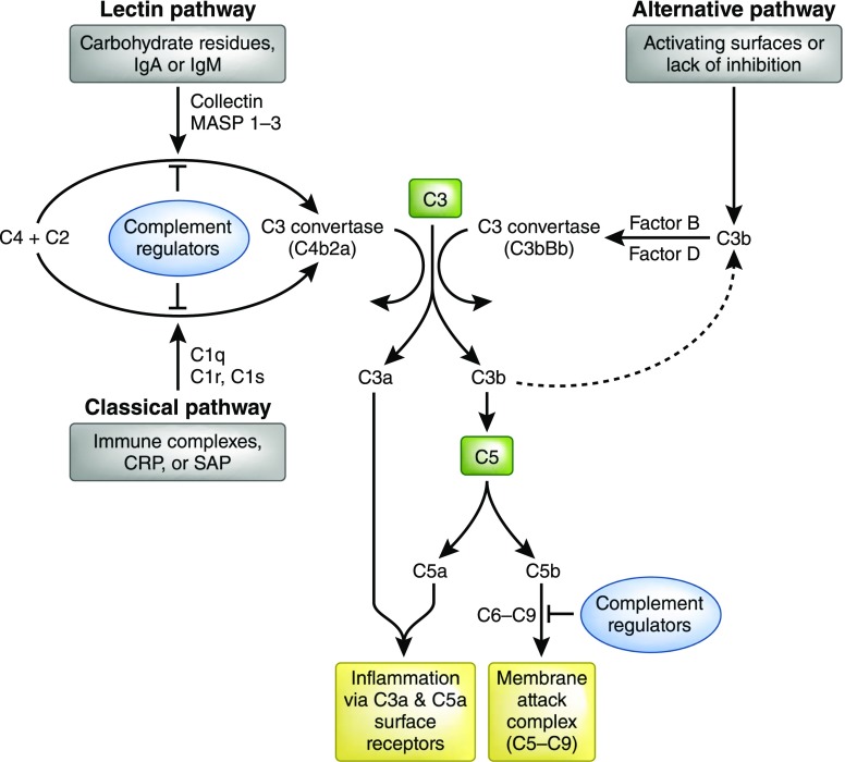
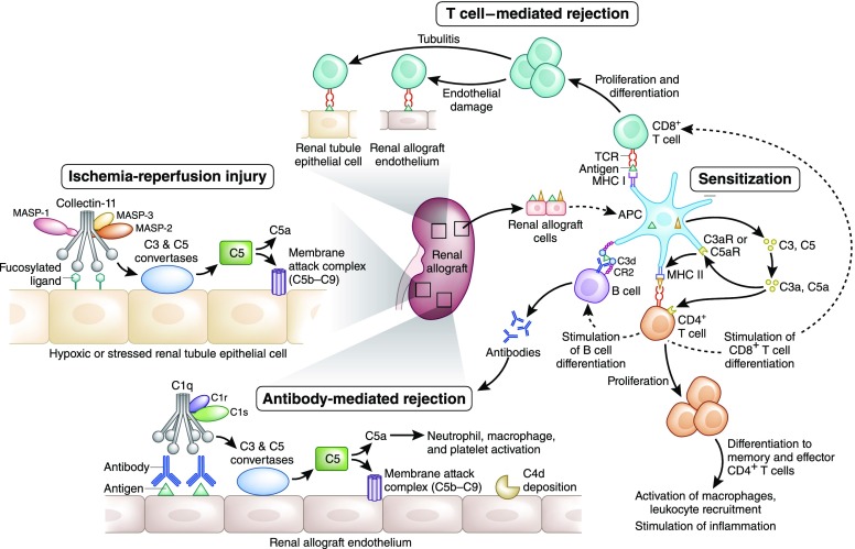
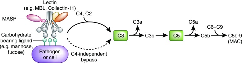
The complement system, consisting of soluble and cell membrane-bound components of the innate immune system, has defined roles in the pathophysiology of renal allograft rejection. Notably, the unavoidable ischemia-reperfusion injury inherent to transplantation is mediated through the terminal complement activation products C5a and C5b-9. Furthermore, biologically active fragments C3a and C5a, produced during complement activation, can modulate both antigen presentation and T cell priming, ultimately leading to allograft rejection. Earlier work identified renal tubule cell synthesis of C3, rather than hepatic synthesis of C3, as the primary source of C3 driving these effects. Recent efforts have focused on identifying the local triggers of complement activation. Collectin-11, a soluble C-type lectin expressed in renal tissue, has been implicated as an important trigger of complement activation in renal tissue. In particular, collectin-11 has been shown to engage L-fucose at sites of ischemic stress, activating the lectin complement pathway and directing the innate immune response to the distressed renal tubule. The interface between collectin-11 and L-fucose, in both the recipient and the allograft, is an attractive target for therapies intended to curtail renal inflammation in the acute phase.

Keywords: collectin-11; complement; lectin pathway; renal transplantation.


Figures

Figure 1.
Figure 1.
The complement cascade. The complement system is activated by one of three major pathways: classical, lectin, or alternative. The classical pathway is triggered by C1 binding to immune surveillance molecules such as IgG, IgM, C-reactive protein (CRP), or serum amyloid protein (SAP) which are attached to the target sequence. The LP is triggered by the binding of collectins, such as MBL and collectin-11, or ficolins to carbohydrate residues on a pathogenic surface or IgA and IgM molecules. The alternative pathway is initiated by direct binding of C3b to activating surfaces. All three pathways converge at the production of the central complement component C3. That is, all pathways form enzyme complexes (classical or alternative convertases) that cleave either C3 (into C3a and C3b) or C5 (into C5a and C5b). C5b triggers the terminal pathway by creating a pore in the target cell membrane via the formation of the membrane attack complex (C5b-C9). Soluble complement effectors C3a and C5a are detected by specific cell receptors thereby promoting inflammation. Complement inhibition occurs via a variety of molecules ultimately inhibiting C3 and C5 convertase or blocking the formation of the membrane attack complex (C5b-C9).
Figure 2.
Figure 2.
Complement activation pathways and allograft immunity. Complement-mediated injury may occur in the intravascular or extravascular compartments. Ischemia-reperfusion injury increases extravascular cell surface expression of a fucosylated ligand that is thought to be recognized by Collectin-11 which associates with MASP-2, in conjunction with MASP-1 and MASP-3, to activate complement via the LP. After the cleavage of C3 and C5, the membrane attack complex (C5b-9) forms, resulting in inflammatory injury and cell death. Complement plays several roles in sensitization against donor alloantigen. Antigen presenting cells (APCs) express complement components C3 and C5 in addition to complement receptors C3aR (C3a Receptor) and C5aR1 (C5a Receptor 1). Generated by complement activation in the extracellular space, C3a and C5a enhance APC priming of T cells by increasing the presentation of alloantigens and the expression of costimulatory molecules. Additionally, C3a and C5a promote CD4+ T cell differentiation and cell longevity. Furthermore, APCs promote proliferation and differentiation of CD4+ and CD8+ T cells. CD8+ T cells mediate cellular rejection in both the intravascular and extravascular compartments which is identified pathologically as endothelitis and tubulitis, respectively. CD4+ T cells stimulate B cell proliferation and ultimately antibody production. In addition, the B cell response to alloantigen may be directly enhanced by complement, because it has been reported for nontransplant antigens, that opsonisation by C3b and its metabolite C3d can enhance antigen presentation via the complement receptor CR2, which is present both on follicular dendritic cells and B cells in secondary lymphoid tissue. Binding of the B cell receptor with the opsonized antigen lowers the threshold for B cell activation and allows for class switching of the donor-specific antibody from IgM to IgG. ABMR occurs when donor-specific antibodies recognize antigens on renal allograft endothelial cells engaging with the C1q, C1r, and C1s complex to initiate complement activation via the classical pathway. Again, C3 and C5 convertases are created and the membrane attack complex subsequently formed. Clinical evidence of complement activation is generally ascribed to the identification of C4d on evaluation of a renal biopsy specimen. All rejection pathways whether complement or cellular based present clinically with evidence of graft dysfunction characterized by increasing serum creatinine and decreasing urine output.
Figure 3.
Figure 3.
The LP. The LP is triggered by collectins, ficolins, and surfactant proteins binding to a carbohydrate moiety such as mannose or fucose that is expressed on a pathogen or stressed cell. The pathway then progresses to formation of C3 convertase and subsequent C3 cleavage which results in terminal complement pathway activation and ultimately formation of C5b-9 (membrane attack complex, MAC). Note that the LP is known to proceed through MASP-2–mediated cleavage of C4 and C2 before formation of C3 convertase. However, recent work has shown the activity of a C4-independent lectin activation pathway leading to cleavage of C3.

References

    1. Beltrame MH, Catarino SJ, Goeldner I, Boldt AB, de Messias-Reason IJ: The lectin pathway of complement and rheumatic heart disease. Front Pediatr 2: 148, 2015 - PMC - PubMed
    1. Sacks SH, Zhou W: The role of complement in the early immune response to transplantation. Nat Rev Immunol 12: 431–442, 2012 - PubMed
    1. Pratt JR, Basheer SA, Sacks SH: Local synthesis of complement component C3 regulates acute renal transplant rejection. Nat Med 8: 582–587, 2002 - PubMed
    1. Loupy A, Lefaucheur C, Vernerey D, Prugger C, Duong van Huyen JP, Mooney N, Suberbielle C, Frémeaux-Bacchi V, Méjean A, Desgrandchamps F, Anglicheau D, Nochy D, Charron D, Empana JP, Delahousse M, Legendre C, Glotz D, Hill GS, Zeevi A, Jouven X: Complement-binding anti-HLA antibodies and kidney-allograft survival. N Engl J Med 369: 1215–1226, 2013 - PubMed
    1. Marsh JE, Farmer CK, Jurcevic S, Wang Y, Carroll MC, Sacks SH: The allogeneic T and B cell response is strongly dependent on complement components C3 and C4. Transplantation 72: 1310–1318, 2001 - PubMed