

Blood Pressure and Heart Rate Measures Associated With Increased Risk of Covert Brain Infarction and Worsening Leukoaraiosis in Older Adults
- PMID: 28663254
- PMCID: PMC5551454
- DOI: 10.1161/ATVBAHA.117.309298
Blood Pressure and Heart Rate Measures Associated With Increased Risk of Covert Brain Infarction and Worsening Leukoaraiosis in Older Adults
Abstract
Objective: In people without previous stroke, covert findings on serial magnetic resonance imaging (MRI) of incident brain infarcts and worsening leukoaraiosis are associated with increased risk for ischemic stroke and dementia. We evaluated whether various measures of blood pressure (BP) and heart rate are associated with these MRI findings.
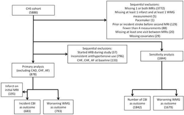
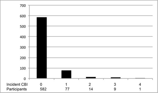
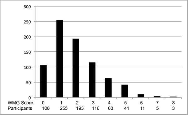
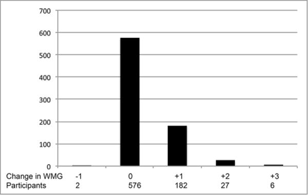
Approach and results: In the CHS (Cardiovascular Health Study), a longitudinal cohort study of older adults, we used relative risk regression to assess the associations of mean, variability, and trend in systolic BP, diastolic BP, and heart rate measured at 4 annual clinic visits between 2 brain MRIs with incident covert brain infarction and worsening white matter grade (using a 10-point scale to characterize leukoaraiosis). We included participants who had both brain MRIs, no stroke before the follow-up MRI, and no change in antihypertensive medication status during follow-up. Among 878 eligible participants, incident covert brain infarction occurred in 15% and worsening white matter grade in 27%. Mean systolic BP was associated with increased risk for incident covert brain infarction (relative risk per 10 mm Hg, 1.28; 95% confidence interval, 1.12-1.47), and mean diastolic BP was associated with increased risk for worsening white matter grade (relative risk per 10 mm Hg, 1.45; 95% confidence interval, 1.24-1.69). These findings persisted in secondary and sensitivity analyses.
Conclusions: Elevated mean systolic BP is associated with increased risk for covert brain infarction, and elevated mean diastolic BP is associated with increased risk for worsening leukoaraiosis. These findings reinforce the importance of hypertension in the development of silent cerebrovascular diseases, but the pathophysiologic relationships to BP for each may differ.
Keywords: blood pressure; heart rate; hypertension; leukoaraiosis; risk factors.
© 2017 American Heart Association, Inc.
Conflict of interest statement
Dr. Psaty serves on the DSMB of a clinical trial funded by the manufacturer (Zoll LifeCor) and on the Steering Committee of the Yale Open Data Access Project funded by Johnson & Johnson.
The remaining authors have no conflicts of interests or relevant financial disclosures.
Figures
Similar articles
-
Left atrial diameter and vascular brain injury on MRI: The Cardiovascular Health Study.Neurology. 2018 Sep 25;91(13):e1237-e1244. doi: 10.1212/WNL.0000000000006228. Epub 2018 Aug 29. Neurology. 2018. PMID: 30158157 Free PMC article.
-
Risk of intraparenchymal hemorrhage with magnetic resonance imaging-defined leukoaraiosis and brain infarcts.Ann Neurol. 2012 Apr;71(4):552-9. doi: 10.1002/ana.22690. Ann Neurol. 2012. PMID: 22522444 Free PMC article.
-
Systolic blood pressure levels among adults with hypertension and incident cardiovascular events: the atherosclerosis risk in communities study.JAMA Intern Med. 2014 Aug;174(8):1252-61. doi: 10.1001/jamainternmed.2014.2482. JAMA Intern Med. 2014. PMID: 24935209 Free PMC article.
-
Incidence, manifestations, and predictors of worsening white matter on serial cranial magnetic resonance imaging in the elderly: the Cardiovascular Health Study.Stroke. 2005 Jan;36(1):56-61. doi: 10.1161/01.STR.0000149625.99732.69. Epub 2004 Nov 29. Stroke. 2005. PMID: 15569873
-
[Blood pressure variation and cardiovascular risk in hypertension].Nihon Rinsho. 2004 Nov;62(11):2145-56. Nihon Rinsho. 2004. PMID: 15552901 Review. Japanese.
Cited by
-
Leukoaraiosis severity is related to increased risk of early neurological deterioration in acute ischemic stroke: a retrospective observational study.Acta Neurol Belg. 2023 Aug;123(4):1413-1420. doi: 10.1007/s13760-023-02249-3. Epub 2023 Apr 4. Acta Neurol Belg. 2023. PMID: 37014516
-
Association Between Blood Pressure Variability and Cerebral Small-Vessel Disease: A Systematic Review and Meta-Analysis.J Am Heart Assoc. 2020 Jan 7;9(1):e013841. doi: 10.1161/JAHA.119.013841. Epub 2019 Dec 24. J Am Heart Assoc. 2020. PMID: 31870233 Free PMC article.
-
Correlation between 24-Hour Ambulatory Blood Pressure Variability and White Matter Lesions in Patients with Cerebral Small Vascular Disease: A Cross-Sectional Study.Comput Math Methods Med. 2022 Aug 8;2022:6364769. doi: 10.1155/2022/6364769. eCollection 2022. Comput Math Methods Med. 2022. Retraction in: Comput Math Methods Med. 2023 Jul 12;2023:9806028. doi: 10.1155/2023/9806028. PMID: 35979055 Free PMC article. Retracted.
-
Electrocardiogram properties and risk of covert brain infarction and other magnetic resonance imaging abnormalities in a stroke-free population.Brain Behav. 2023 May;13(5):e2991. doi: 10.1002/brb3.2991. Epub 2023 Apr 16. Brain Behav. 2023. PMID: 37062924 Free PMC article.
-
Blood Pressure Variability and Cerebral Small Vessel Disease: A Systematic Review and Meta-Analysis of Population-Based Cohorts.Stroke. 2020 Jan;51(1):82-89. doi: 10.1161/STROKEAHA.119.026739. Epub 2019 Nov 27. Stroke. 2020. PMID: 31771460 Free PMC article.
References
-
- Fanning JP, Wesley AJ, Wong AA, Fraser JF. Emerging spectra of silent brain infarction. Stroke. 2014;45:3461–3471. - PubMed
-
- Vermeer SE, Longstreth WT, Jr, Koudstaal PJ. Silent brain infarcts: a systematic review. Lancet Neurol. 2007;6:611–619. - PubMed
-
- Price TR, Manolio TA, Kronmal RA, Kittner SJ, Yue NC, Robbins J, Anton-Culver H, O’Leary DH. Silent brain infarction on magnetic resonance imaging and neurological abnormalities in community-dwelling older adults. The Cardiovascular Health Study. Stroke. 1997;28:1158–1164. - PubMed
Publication types
MeSH terms
Substances
Grants and funding
- N01 HC085080/HL/NHLBI NIH HHS/United States
- U01 HL080295/HL/NHLBI NIH HHS/United States
- N01 HC085082/HL/NHLBI NIH HHS/United States
- U01 HL130114/HL/NHLBI NIH HHS/United States
- HHSN268201200036C/HL/NHLBI NIH HHS/United States
- N01 HC055222/HL/NHLBI NIH HHS/United States
- N01 HC085079/HL/NHLBI NIH HHS/United States
- R01 AG023629/AG/NIA NIH HHS/United States
- N01 HC085081/HL/NHLBI NIH HHS/United States
- HHSN268200800007C/HL/NHLBI NIH HHS/United States
- N01 HC015103/HC/NHLBI NIH HHS/United States
- N01 HC085086/HL/NHLBI NIH HHS/United States
- N01 HC085083/HL/NHLBI NIH HHS/United States
LinkOut - more resources
Full Text Sources
Other Literature Sources
Medical
