Smooth Muscle Cells Derived From Second Heart Field and Cardiac Neural Crest Reside in Spatially Distinct Domains in the Media of the Ascending Aorta-Brief Report
- PMID: 28663257
- PMCID: PMC5570666
- DOI: 10.1161/ATVBAHA.117.309599
Smooth Muscle Cells Derived From Second Heart Field and Cardiac Neural Crest Reside in Spatially Distinct Domains in the Media of the Ascending Aorta-Brief Report
Abstract
Objective: Smooth muscle cells (SMCs) of the proximal thoracic aorta are embryonically derived from the second heart field (SHF) and cardiac neural crest (CNC). However, distributions of these embryonic origins are not fully defined. The regional distribution of SMCs of different origins is speculated to cause region-specific aortopathies. Therefore, the aim of this study was to determine the distribution of SMCs of SHF and CNC origins in the proximal thoracic aorta.
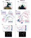
Approach and results: Mice with repressed LacZ in the ROSA26 locus were bred to those expressing Cre controlled by either the Wnt1 or Mef2c (myocyte-specific enhancer factor 2c) promoter to trace CNC- and SHF-derived SMCs, respectively. Thoracic aortas were harvested, and activity of β-galactosidase was determined. Aortas from Wnt1-Cre mice had β-galactosidase-positive areas throughout the region from the proximal ascending aorta to just distal of the subclavian arterial branch. Unexpectedly, β-galactosidase-positive areas in Mef2c-Cre mice extended from the aortic root throughout the ascending aorta. This distribution occurred independent of sex and aging. Cross and sagittal aortic sections demonstrated that CNC-derived cells populated the inner medial aspect of the anterior region of the ascending aorta and transmurally in the media of the posterior region. Interestingly, outer medial cells throughout anterior and posterior ascending aortas were derived from the SHF. β-Galactosidase-positive medial cells of both origins colocalized with an SMC marker, α-actin.
Conclusions: Both CNC- and SHF-derived SMCs populate the media throughout the ascending aorta. The outer medial cells of the ascending aorta form a sleeve populated by SHF-derived SMCs.
Keywords: aorta; cell tracking; neural crest; second heart field; smooth muscle.
© 2017 American Heart Association, Inc.
Conflict of interest statement
Figures


Similar articles
-
Single-Cell Transcriptomics Identifies Selective Lineage-Specific Regulation of Genes in Aortic Smooth Muscle Cells in Mice.Arterioscler Thromb Vasc Biol. 2025 Feb;45(2):e15-e29. doi: 10.1161/ATVBAHA.124.321482. Epub 2025 Jan 2. Arterioscler Thromb Vasc Biol. 2025. PMID: 39744838
-
Heterogeneous Cardiac-Derived and Neural Crest-Derived Aortic Smooth Muscle Cells Exhibit Similar Transcriptional Changes After TGFβ Signaling Disruption.Arterioscler Thromb Vasc Biol. 2025 Feb;45(2):260-276. doi: 10.1161/ATVBAHA.124.321706. Epub 2024 Dec 19. Arterioscler Thromb Vasc Biol. 2025. PMID: 39697172
-
Smooth muscle cells and fibroblasts in the ascending aorta exhibit minor differences between embryonic origins in angiotensin II-driven transcriptional alterations.Sci Rep. 2025 May 13;15(1):16617. doi: 10.1038/s41598-025-99862-4. Sci Rep. 2025. PMID: 40360730 Free PMC article.
-
Embryonic Heterogeneity of Smooth Muscle Cells in the Complex Mechanisms of Thoracic Aortic Aneurysms.Genes (Basel). 2022 Sep 9;13(9):1618. doi: 10.3390/genes13091618. Genes (Basel). 2022. PMID: 36140786 Free PMC article. Review.
-
Cardiac Neural Crest.Cold Spring Harb Perspect Biol. 2021 Jan 4;13(1):a036715. doi: 10.1101/cshperspect.a036715. Cold Spring Harb Perspect Biol. 2021. PMID: 32071091 Free PMC article. Review.
Cited by
-
Generation of Vascular Smooth Muscle Cells From Induced Pluripotent Stem Cells: Methods, Applications, and Considerations.Circ Res. 2021 Mar 5;128(5):670-686. doi: 10.1161/CIRCRESAHA.120.318049. Epub 2021 Mar 4. Circ Res. 2021. PMID: 33818124 Free PMC article. Review.
-
HDAC1-mediated repression of the retinoic acid-responsive gene ripply3 promotes second heart field development.PLoS Genet. 2019 May 15;15(5):e1008165. doi: 10.1371/journal.pgen.1008165. eCollection 2019 May. PLoS Genet. 2019. PMID: 31091225 Free PMC article.
-
Vascular Development.Arterioscler Thromb Vasc Biol. 2018 Mar;38(3):e17-e24. doi: 10.1161/ATVBAHA.118.310223. Arterioscler Thromb Vasc Biol. 2018. PMID: 29467221 Free PMC article. Review.
-
Is Transposition of the Great Arteries Associated With Shortening of the Intrapericardial Portions of the Great Arterial Trunks? An Echocardiographic Analysis on Newborn Infants With Simple Transposition of the Great Arteries to Explore an Animal Model-Based Hypothesis on Human Beings.J Am Heart Assoc. 2021 Aug 3;10(15):e019334. doi: 10.1161/JAHA.120.019334. Epub 2021 Jul 19. J Am Heart Assoc. 2021. PMID: 34278802 Free PMC article.
-
Notch1 haploinsufficiency causes ascending aortic aneurysms in mice.JCI Insight. 2017 Nov 2;2(21):e91353. doi: 10.1172/jci.insight.91353. JCI Insight. 2017. PMID: 29093270 Free PMC article.
References
-
- Hiratzka LF, Bakris GL, Beckman JA, et al. 2010 ACCF/AHA/AATS/ACR/ASA/ SCA/ SCAI/SIR/STS/SVM guidelines for the diagnosis and management of patients with Thoracic Aortic Disease. Circulation. 2010;121:e266–369. - PubMed
-
- Osada H, Kyogoku M, Ishidou M, Morishima M, Nakajima H. Aortic dissection in the outer third of the media: what is the role of the vasa vasorum in the triggering process? Eur J Cardiothorac Surg. 2013;43:e82–88. - PubMed
-
- Majesky MW. Developmental basis of vascular smooth muscle diversity. Arterioscler Thromb Vasc Biol. 2007;27:1248–1258. - PubMed
-
- Le Lievre CS, Le Douarin NM. Mesenchymal derivatives of the neural crest: analysis of chimaeric quail and chick embryos. J Embryol Exp Morphol. 1975;34:125–154. - PubMed
Publication types
MeSH terms
Substances
Grants and funding
LinkOut - more resources
Full Text Sources
Other Literature Sources
Molecular Biology Databases

