How Common Is Signal-Intensity Increase in Optic Nerve Segments on 3D Double Inversion Recovery Sequences in Visually Asymptomatic Patients with Multiple Sclerosis?
- PMID: 28663263
- PMCID: PMC7963692
- DOI: 10.3174/ajnr.A5262
How Common Is Signal-Intensity Increase in Optic Nerve Segments on 3D Double Inversion Recovery Sequences in Visually Asymptomatic Patients with Multiple Sclerosis?
Abstract
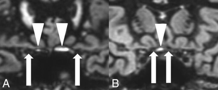
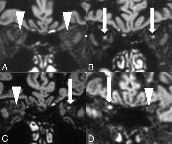
Background and purpose: In postmortem studies, subclinical optic nerve demyelination is very common in patients with MS but radiologic demonstration is difficult and mainly based on STIR T2WI. Our aim was to evaluate 3D double inversion recovery MR imaging for the detection of subclinical demyelinating lesions within optic nerve segments.
Materials and methods: The signal intensities in 4 different optic nerve segments (ie, retrobulbar, canalicular, prechiasmatic, and chiasm) were evaluated on 3D double inversion recovery MR imaging in 95 patients with MS without visual symptoms within the past 3 years and in 50 patients without optic nerve pathology. We compared the signal intensities with those of the adjacent lateral rectus muscle. The evaluation was performed by a student group and an expert neuroradiologist. Statistical evaluation (the Cohen κ test) was performed.
Results: On the 3D double inversion recovery sequence, optic nerve segments in the comparison group were all hypointense, and an isointense nerve sheath surrounded the retrobulbar nerve segment. At least 1 optic nerve segment was isointense or hyperintense in 68 patients (72%) in the group with MS on the basis of the results of the expert neuroradiologist. Student raters were able to correctly identify optic nerve hypersignal in 97%.
Conclusions: A hypersignal in at least 1 optic nerve segment on the 3D double inversion recovery sequence compared with hyposignal in optic nerve segments in the comparison group was very common in visually asymptomatic patients with MS. The signal-intensity rating of optic nerve segments could also be performed by inexperienced student readers.
© 2017 by American Journal of Neuroradiology.
Figures


Similar articles
-
Detecting optic nerve lesions in clinically isolated syndrome and multiple sclerosis: double-inversion recovery magnetic resonance imaging in comparison with visually evoked potentials.J Neurol. 2019 Jan;266(1):148-156. doi: 10.1007/s00415-018-9114-2. Epub 2018 Nov 16. J Neurol. 2019. PMID: 30446963
-
Detecting optic nerve lesions in multiple sclerosis patients with a 1,5 T MRI: Evaluation of a 3D DIR sequence compared to a 2D STIR sequence.Mult Scler Relat Disord. 2024 Oct;90:105832. doi: 10.1016/j.msard.2024.105832. Epub 2024 Aug 24. Mult Scler Relat Disord. 2024. PMID: 39213862
-
Asymptomatic optic nerve lesions: An underestimated cause of silent retinal atrophy in MS.Neurology. 2020 Jun 9;94(23):e2468-e2478. doi: 10.1212/WNL.0000000000009504. Epub 2020 May 20. Neurology. 2020. PMID: 32434868
-
Three-dimensional MRI sequences in MS diagnosis and research.Mult Scler. 2019 Nov;25(13):1700-1709. doi: 10.1177/1352458519848100. Epub 2019 May 22. Mult Scler. 2019. PMID: 31116071 Review.
-
3D double inversion recovery MR imaging: Clinical applications and usefulness in a wide spectrum of central nervous system diseases.J Neuroradiol. 2019 Mar;46(2):107-116. doi: 10.1016/j.neurad.2018.06.002. Epub 2018 Jul 25. J Neuroradiol. 2019. PMID: 30016704 Review.
Cited by
-
Detecting optic nerve lesions in clinically isolated syndrome and multiple sclerosis: double-inversion recovery magnetic resonance imaging in comparison with visually evoked potentials.J Neurol. 2019 Jan;266(1):148-156. doi: 10.1007/s00415-018-9114-2. Epub 2018 Nov 16. J Neurol. 2019. PMID: 30446963
-
Evaluating the Utility of a Postprocessing Algorithm for MRI Evaluation of Optic Neuritis.AJNR Am J Neuroradiol. 2019 Jun;40(6):1043-1048. doi: 10.3174/ajnr.A6057. Epub 2019 May 2. AJNR Am J Neuroradiol. 2019. PMID: 31048299 Free PMC article.
-
Optic Nerve Lesion Length at the Acute Phase of Optic Neuritis Is Predictive of Retinal Neuronal Loss.Neurol Neuroimmunol Neuroinflamm. 2022 Jan 28;9(2):e1135. doi: 10.1212/NXI.0000000000001135. Print 2022 Mar. Neurol Neuroimmunol Neuroinflamm. 2022. PMID: 35091465 Free PMC article. Clinical Trial.
-
Long segment 3D double inversion recovery (DIR) hypersignal on MRI in glaucomatous optic neuropathy.BMC Ophthalmol. 2019 Dec 16;19(1):258. doi: 10.1186/s12886-019-1273-0. BMC Ophthalmol. 2019. PMID: 31842814 Free PMC article.
-
Improved detection of multiple sclerosis lesions with T2-prepared double inversion recovery at 3T.J Neuroimaging. 2022 Sep;32(5):902-909. doi: 10.1111/jon.13021. Epub 2022 Jul 1. J Neuroimaging. 2022. PMID: 35776654 Free PMC article.
References
MeSH terms
LinkOut - more resources
Full Text Sources
Other Literature Sources
Medical
Miscellaneous