

The asbestos fibre burden in human lungs: new insights into the chrysotile debate
- PMID: 28663314
- PMCID: PMC5898940
- DOI: 10.1183/13993003.02534-2016
The asbestos fibre burden in human lungs: new insights into the chrysotile debate
Abstract
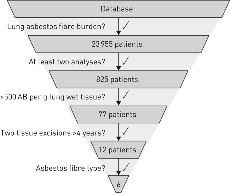
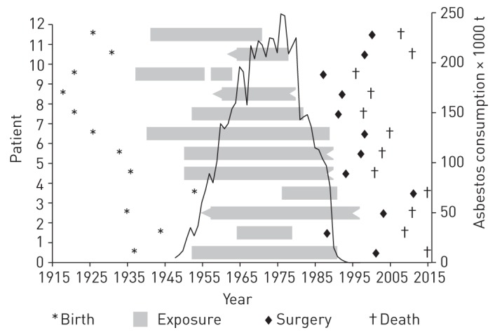
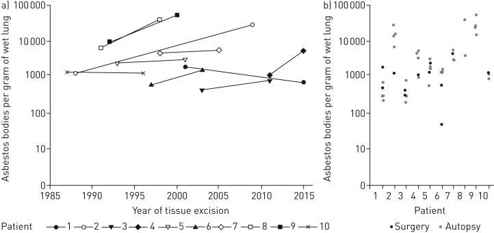
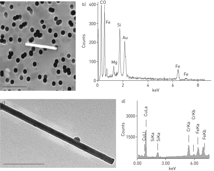
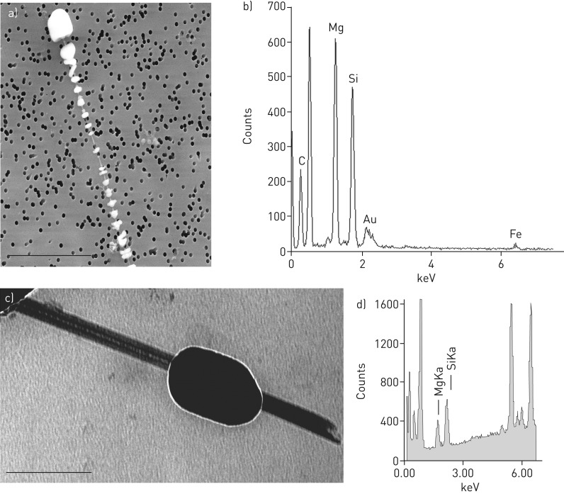
The traceability of asbestos fibres in human lungs is a matter of discussion especially for chrysotile. This issue is of high significance for differential diagnosis, risk assessment and occupational compensation. At present no intra-individual longitudinal information is available. This study addresses the question whether the asbestos fibre burden in human lungs decreases with time after exposure cessation.The database of the German Mesothelioma Register was screened for patients with asbestos body counts of at least 500 fibres per gram of wet lung, which had been analysed twice from different tissue excisions at minimum intervals of 4 years.Twelve datasets with individual longitudinal information were discovered with a median interval of about 8 years (range 4-21 years). Both examinations were performed after exposure cessation (median: surgery, 9.5 years; autopsy, 22 years). Pulmonary asbestos fibre burden was stable between both examinations (median 1623/4269 asbestos bodies per gram wet lung). Electron microscopy demonstrated a preponderance of chrysotile (median 80%).This study is the first to present longitudinal intra-individual data about the asbestos fibre burden in living human lungs. The high biopersistence of amphiboles, but also of chrysotile, offers mechanistic explanations for fibre toxicity, especially the long latency period of asbestos-related diseases.
Copyright ©ERS 2017.
Conflict of interest statement
Conflict of interest: Disclosures can be found alongside this article at erj.ersjournals.com
Figures
Comment in
-
Quantifying asbestos in lung tissue: what debate?Eur Respir J. 2017 Jun 29;49(6):1700861. doi: 10.1183/13993003.00861-2017. Print 2017 Jun. Eur Respir J. 2017. PMID: 28663320 No abstract available.
-
Correspondence regarding the article "The asbestos fibre burden in human lungs: new insights into the chrysotile debate".Eur Respir J. 2017 Dec 21;50(6):1701644. doi: 10.1183/13993003.01644-2017. Print 2017 Dec. Eur Respir J. 2017. PMID: 29269580 No abstract available.
-
Correspondence regarding the article "The asbestos fibre burden in human lungs: new insights into the chrysotile debate".Eur Respir J. 2017 Dec 21;50(6):1702188. doi: 10.1183/13993003.02188-2017. Print 2017 Dec. Eur Respir J. 2017. PMID: 29269586 No abstract available.
-
Correspondence regarding the article "The asbestos fibre burden in human lungs: new insights into the chrysotile debate".Eur Respir J. 2017 Dec 21;50(6):1702204. doi: 10.1183/13993003.02204-2017. Print 2017 Dec. Eur Respir J. 2017. PMID: 29269588 No abstract available.
Similar articles
-
Lung fibre burden and risk of malignant mesothelioma in shipyard workers: a necropsy-based case-control study.Ann Work Expo Health. 2024 Jun 6;68(5):476-485. doi: 10.1093/annweh/wxae018. Ann Work Expo Health. 2024. PMID: 38532179
-
Inorganic fibres in the lung tissue of Hungarian and German lung cancer patients.Int Arch Occup Environ Health. 2001 Mar;74(2):133-8. doi: 10.1007/s004200000202. Int Arch Occup Environ Health. 2001. PMID: 11317707
-
Biological indicators of chrysotile exposure.Ann Occup Hyg. 1994 Aug;38(4):503-18, 410-1. doi: 10.1093/annhyg/38.4.503. Ann Occup Hyg. 1994. PMID: 7978972
-
Chrysotile asbestos is the main cause of pleural mesothelioma.Am J Ind Med. 1996 Sep;30(3):252-66. doi: 10.1002/(SICI)1097-0274(199609)30:3<252::AID-AJIM2>3.0.CO;2-0. Am J Ind Med. 1996. PMID: 8876792 Review.
-
Deposition and retention of inhaled fibres: effects on incidence of lung cancer and mesothelioma.Occup Environ Med. 1994 Dec;51(12):793-8. doi: 10.1136/oem.51.12.793. Occup Environ Med. 1994. PMID: 7849861 Free PMC article. Review.
Cited by
-
An Emerging Model for Cancer Development from a Tumor Microenvironment Perspective in Mice and Humans.Adv Exp Med Biol. 2020;1225:19-29. doi: 10.1007/978-3-030-35727-6_2. Adv Exp Med Biol. 2020. PMID: 32030645 Review.
-
Prevention of Asbestos Exposure in Latin America within a Global Public Health Perspective.Ann Glob Health. 2019 Mar 29;85(1):49. doi: 10.5334/aogh.2341. Ann Glob Health. 2019. PMID: 30924615 Free PMC article. Review.
-
Amphibole asbestos as an environmental trigger for systemic autoimmune diseases.Autoimmun Rev. 2024 Jul-Aug;23(7-8):103603. doi: 10.1016/j.autrev.2024.103603. Epub 2024 Aug 20. Autoimmun Rev. 2024. PMID: 39154740 Free PMC article. Review.
-
Asbestos Bodies Burden in the Autopsy Lung Tissue from General Thai Population.Open Respir Med J. 2019 May 31;13:5-10. doi: 10.2174/1874306401913010005. eCollection 2019. Open Respir Med J. 2019. PMID: 31929836 Free PMC article.
-
The incidence of malignancies in asbestosis with chrysotile exposure: a large Chinese prospective cohort study.Front Oncol. 2023 Jul 6;13:1172496. doi: 10.3389/fonc.2023.1172496. eCollection 2023. Front Oncol. 2023. PMID: 37483507 Free PMC article.
References
-
- Yusa T, Hiroshima K, Sakai F, et al. . Significant relationship between the extent of pleural plaques and pulmonary asbestos body concentration in lung cancer patients with occupational asbestos exposure. Am J Ind Med 2015; 58: 444–455. - PubMed
-
- Attanoos RL, Alchami FS, Pooley FD, et al. . Usual interstitial pneumonia in asbestos-exposed cohorts – concurrent idiopathic pulmonary fibrosis or atypical asbestosis? Histopathology 2016; 69: 492–498. - PubMed
-
- Henderson D, Rantanen J, Barnhart S, et al. . Asbestos, asbestosis and cancer: the Helsinki criteria for diagnosis and attribution. Scand J Work Environ Health 1997; 23: 311–316. - PubMed
Publication types
MeSH terms
Substances
LinkOut - more resources
Full Text Sources
Other Literature Sources
Medical